Brain Diseases
Cavernoma (Cavernous Malformation)
Back to Brain DiseasesWhat are Cavernomas?
Cavernomas, also known as cavernous malformations, cavernous angiomas, or cavernous hemangiomas, are abnormal clusters of tightly packed small blood vessels, or capillaries. While they can occur throughout the body, they can cause problems if they form in the brain, the brainstem, or (in rare cases) in the spinal cord. They are not tumors or compressions, but the walls of these blood vessel malformations tend to be extremely thin, and cavernomas can rupture or bleed, causing a variety of neurologic symptoms and potentially seizures or strokes.

20%
of cavernomas develop due to genetic factors.
20 to 40 years
is the typical age for the onset of symptoms.
1 in 500
people have at least one cavernoma in their brain.
Cavernoma Risk Factors
Cavernoma Signs and Symptoms
Cavernoma Diagnosis
Cavernoma Treatments
Cavernoma Research Efforts
Cavernous Malformation Resources
Cleveland Clinic: Cavernous Malformation (Cavernoma)
Mayo Clinic: Cavernous Malformations
Alliance to Cure Cavernous Malformations

Support Brain Research
We need your help to continue to fund brain research projects and find cures. Stand with us in the fight against brain diseases and disorders.
Additional Brain Disease Information
Explore more brain disease information, research, and stories.
BLOG
News and Articles
Discover the latest news in brain disease research, hear stories from people affected by brain disease and their caregivers, read up on brain disease-specific information, and more.














Rapid Eye Movement Behavior Sleep Disorder: One Person’s Journey to Delay Neurodegenerative Disease
Read More


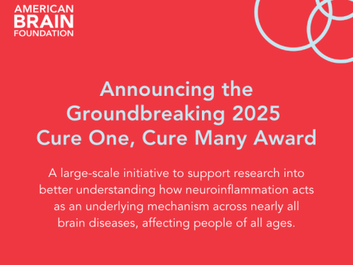
$10 Million Research Initiative Established to Understand Neuroinflammation’s Impact on Brain Diseases
Read More




How Spinal Muscular Atrophy Research Is Fueling Advancements for Other Neuromuscular Diseases
Read More





Cerebral Small Vessel Disease: Learn About One Person’s Story of Early Dementia, Misdiagnosis, and Living With CSVD
Read More
Featured Researcher: Dr. Carolina Barnett-Tapia, MD, PhD, Institute of Health Policy, Management and Evaluation, University of Ontario
Read More

Researcher Feature: Jasmeer Chhatwal, MD, PhD, Harvard Medical School and Massachusetts General Hospital
Read More





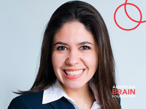
Featured Researcher: Lidia Maria Moura, MD, MPH, PhD, Center for Value-Based Health Care and Sciences at Massachusetts General Hospital
Read More






Stephen L. Hauser, MD: Career-Long Dedication Leads to Breakthrough Research in Multiple Sclerosis
Read More
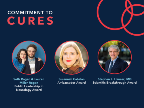
Seth Rogen and Lauren Miller Rogen to Accept Award at Commitment to Cures on April 6 in Support of Brain Disease Research
Read More
Announcing the 2022 Ted M. Burns Humanism in Neurology Award Recipient: Barbara S. Giesser, MD, FAAN
Read More
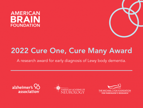
Recipients Selected for the American Brain Foundation 2022 Cure One, Cure Many Award for the Early Diagnosis of Lewy Body Dementia
Read More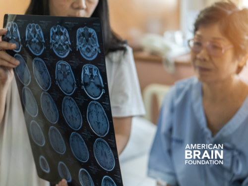


Prioritizing Healthcare Disparities Research: Groundbreaking American Brain Foundation-Funded Awards
Read More





Groundbreaking research award established to discover an accurate method of diagnosing Lewy body dementia
Read More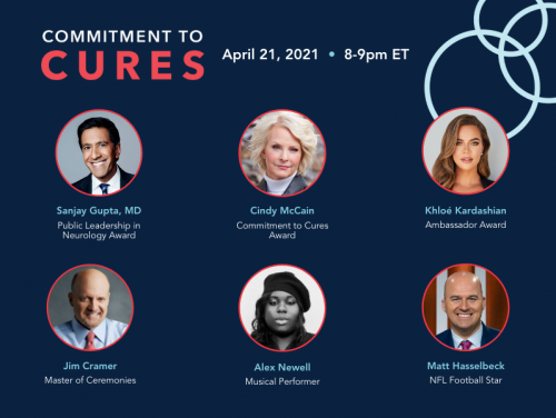
Star-Studded Virtual Event to Recognize Breakthroughs in Brain Disease Research on April 21
Read More









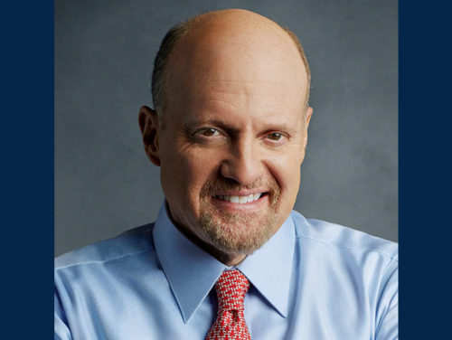
Jim Cramer to Receive American Brain Foundation’s 2020 Ambassador Award for Migraine Advocacy
Read More
American Brain Foundation Gala in Toronto to Recognize Breakthroughs in Brain Disease Research, Celebrate Advocates
Read More





International Sponsor Council Focuses on Brain Health as Part of Its Athlete Health Initiative
Read More
Foundation to present prestigious Public Leadership in Neurology Award to B. Smith and Dan Gasby
Read More
Orlena
Orlena was driving with her young daughter in the car when she suffered a stroke because of a rare brain vessel disorder called Moyamoya. After two brain surgeries and six months unable to care for her daughter, Orlena began to reclaim her independence and became a Moyamoya advocate.
Read More of Orlena’s Story
ABF Stories
Orlena
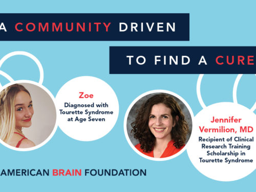
Zoe
At 15 years old, Zoe has been navigating her Tourette syndrome diagnosis for eight years. “I had to figure out at a young age how my emotions affect my tics, from good to awful ways. It has been frustrating to figure out on my own and learn to deal with. This was the first moment in my life where my parents couldn’t fix something for me.”
Read More of Zoe’s Story
ABF STORIES
Zoe
Sasha
Sasha was diagnosed with epilepsy after sustaining a soccer-related brain injury in high school. Since then, Sasha has worked to find both a treatment that controlled her seizures and a career that fulfilled her desire to care for others. “Life isn’t meant to be lived taking multiple medications and having seizures you can’t control.”
Read More of Sasha’s Story
ABF STORIES
Sasha
Ken Jr.
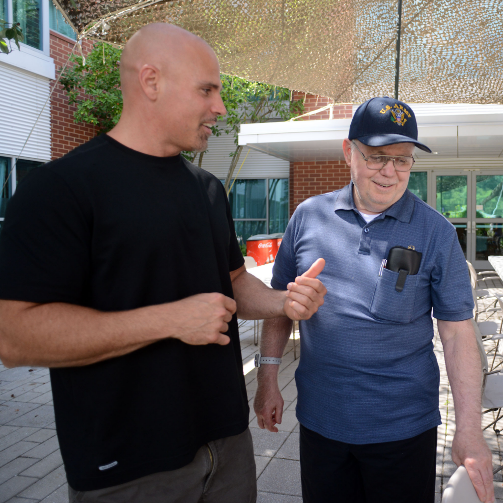
Before frontotemporal lobe dementia, sometimes called Pick’s disease, Ken Keene Sr. was a respected community member and a handyman who could fix anything. For his oldest son and namesake Ken Keene Jr., there was nothing his father couldn’t do: “To my family, he was just that super dad. Then all of a sudden he’s reaching out for help.”
Read More of Ken’s Story
ABF STORIES
Ken Jr.
Morgan
It started with a twinge. After a simple surgery and some physical therapy, Morgan wasn’t getting better. It took two more surgeries before she was diagnosed with Complex Regional Pain Syndrome (CRPS) type II.
Read More of Morgan’s Story
ABF STORIES
Morgan
Kelly
In 2018, Kelly was diagnosed with glioblastoma after an MRI confirmed that a golf-ball-sized tumor above her right ear was affecting the left side of her body. After surgery to remove the tumor, Kelly was able to get through her darkest days with a healthy dose of humor. Now she is an advocate for other patients living with strokes and brain tumors.
Read More of Kelly’s Story
ABF STORIES
Kelly
Nancy
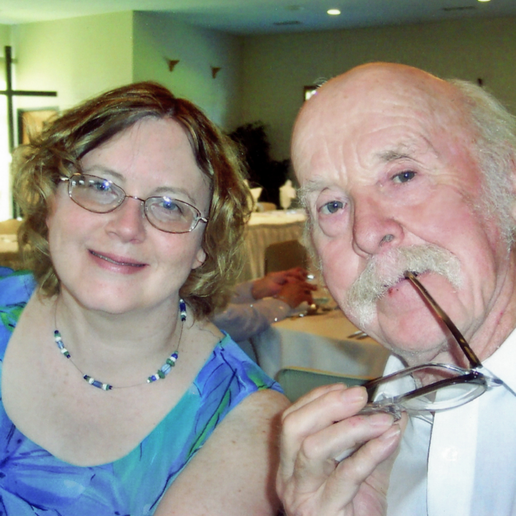
When her father was diagnosed with vascular dementia in 2005, Nancy quickly shifted into the role of his caregiver. Nancy and her sisters drew from their individual strengths to provide their father with medical assistance and emotional support until his passing in 2012. At that time, he was diagnosed again with Lewy body dementia. “No matter what your financial situation, there’s probably going to be help out there… Just don’t do it alone, because burning yourself out is not going to help anybody.”
Read More of Nancy’s Story
ABF STORIES
Nancy
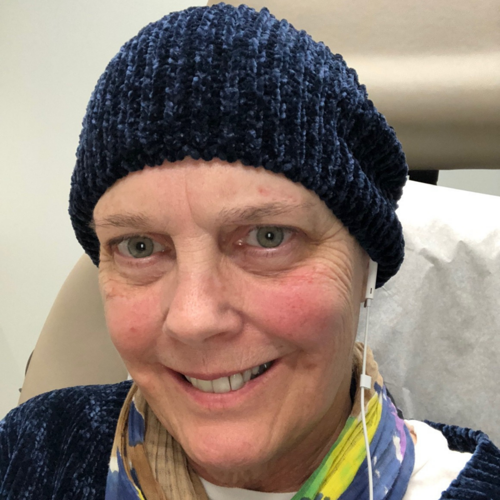
Maureen
Maureen worked as a high school English teacher until she started experiencing a confusing array of symptoms that affected her ability to work, along with her relationships with family and friends. After visiting numerous specialists for years, Maureen received a delayed diagnosis of Lewy body dementia. Now Maureen wants to raise awareness about what life is like with living with this disease. “I felt that I just wanted to offer something to this community that I hadn’t been able to find really.”
Read More of Maureen’s Story
ABF STORIES
Maureen
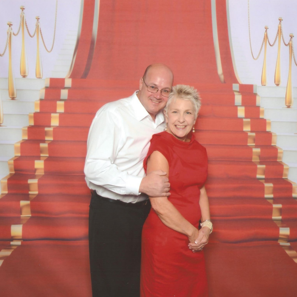
Joey
For the first 32 years of his life, Joey experienced seizures from epilepsy. For several years, Joey worked hard in his career as a pipe welder and in construction until his seizures started happening more frequently and intensely. Brain surgery became Joey’s only viable option for treatment. Doctors were unsure if Joey would be able to survive after finding more brain damage than expected, but he surprised everyone by thriving during his post-surgery recovery.
Read More of Joey’s Story
ABF STORIES
Joey
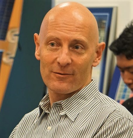
Ben
At age 40, Ben was perfectly healthy and active until he started noticing the early symptoms of a neurological condition that weakened his physical movements. After a 2-year search for his diagnosis of X-linked adrenoleukodystrophy, he began his journey as an advocate for others living with rare diseases. Last year, Ben joined the board of the American Brain Foundation to further his impact.
Read More of Ben’s Story
ABF STORIES
Ben
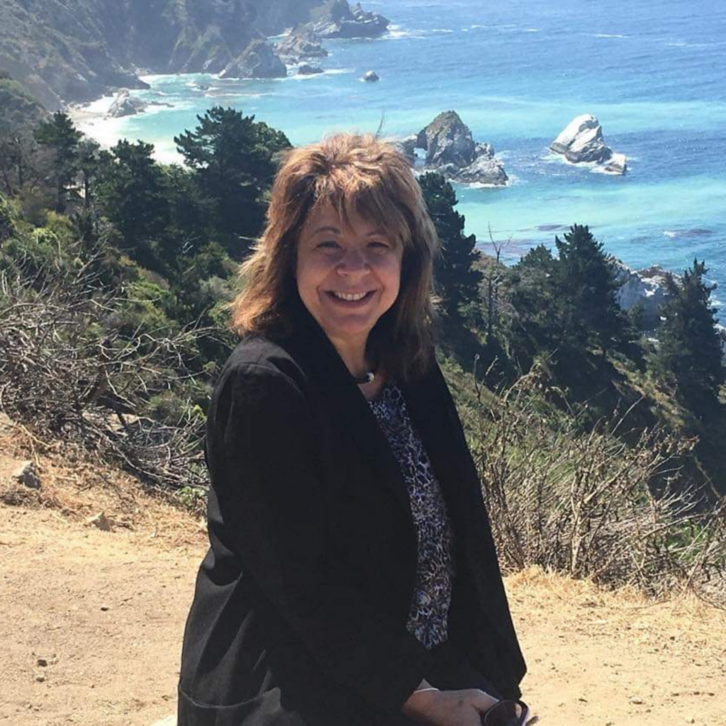
Michele
In 2002, while her young daughter battled a fatal diagnosis of pons glioma, Michele experienced a life-threatening brain hemorrhage when a brain malformation she likely had since birth ruptured. She experienced drastic changes in the way her brain functioned, especially in regard to memory and spatial awareness. Today, Michele is still learning to adjust. “When someone’s brain is not functioning correctly it impacts everything in their life. It impacts how they think, how they feel about themselves, it’s a very hopeless space to be in as a person.”
Read More of Michele’s Story
ABF STORIES
Michele
Matt
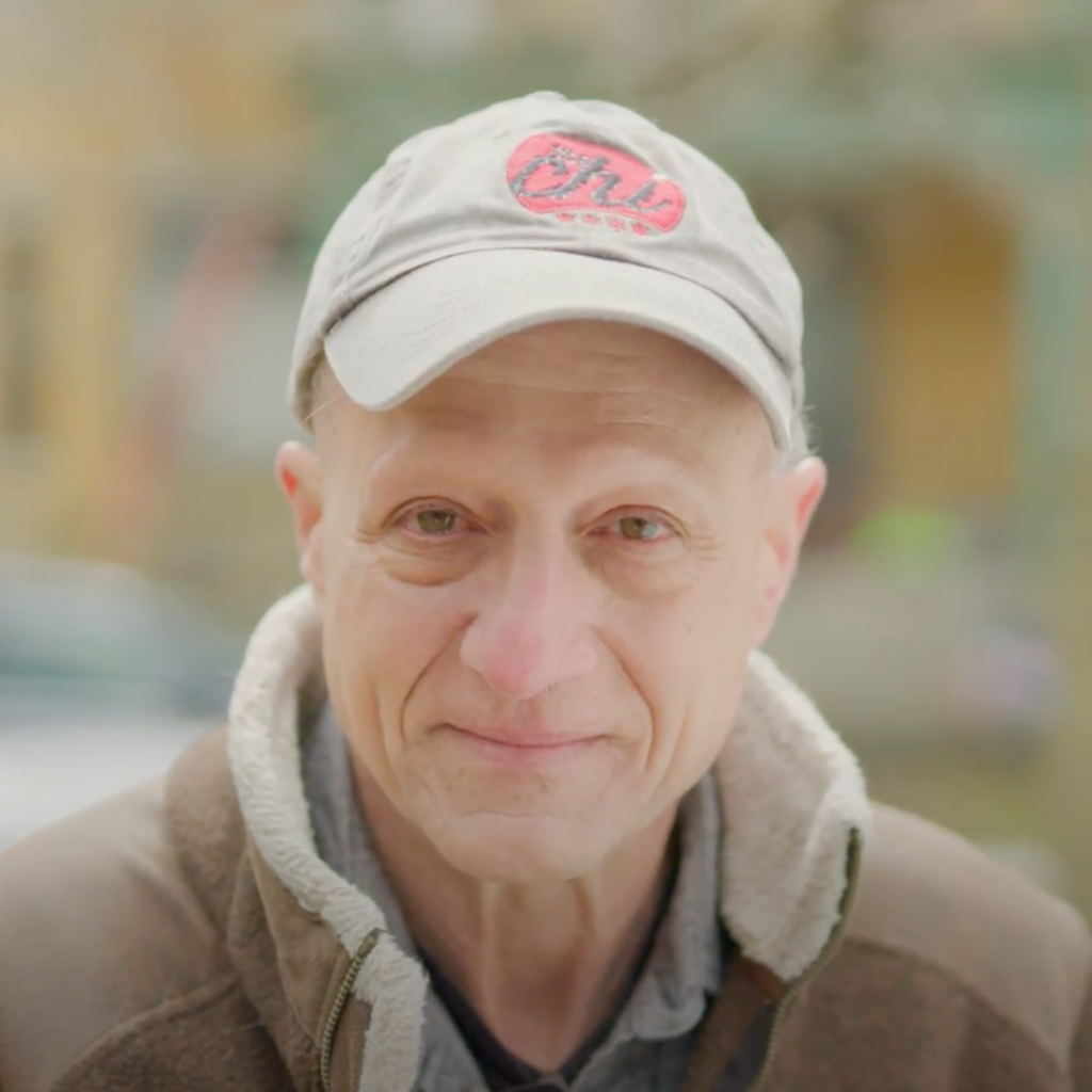
Matt is a husband, father, and professional photographer. Over the past 11 years, he has had surgeries and radiation to remove five benign brain tumors. Following the third surgery, he began to experience more struggles in his daily life. “I think the biggest loss is my relationship to my family. They saw me as the dad and strong. Now they can’t count on me as much. It’s important to support people with brain disease ‘cause it can happen to anybody.’”
Read More of Matt’s Story
ABF STORIES
Matt
Mary Jo
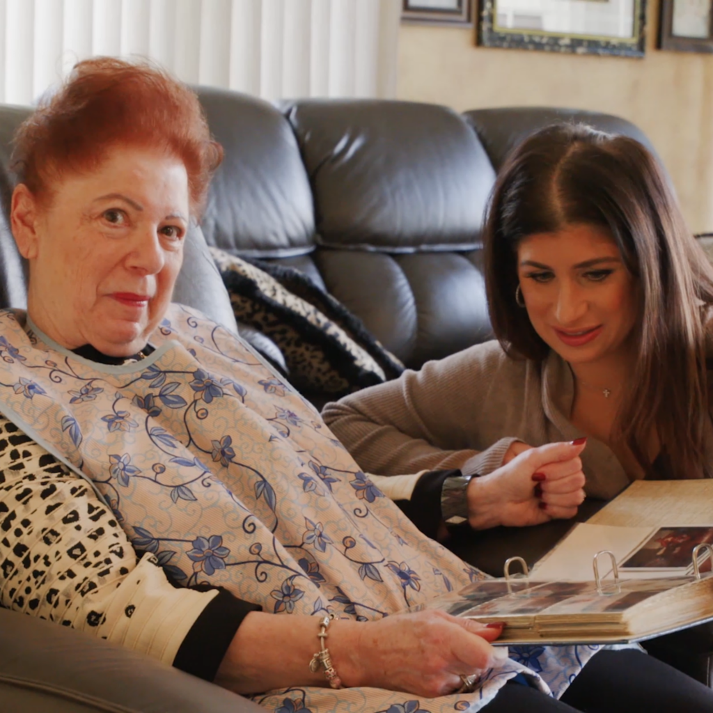
When she was in her early 30’s, Mary Jo’s relationship with her parents was forever altered by brain disease. Her father suffered a subdural hematoma, was diagnosed with diabetic neuropathy, and experienced a stroke while her mother was diagnosed with Alzheimer’s and dementia. “They’re a responsibility that I have to take care of as they have taken care of me.”
Read More of Mary Jo’s Story
ABF STORIES
Mary Jo
Tom
Tom suffers from dystonia, a painful neurological movement disorder with no known cure. When he first started experiencing strange pains and cramps in his neck muscles, he saw chiropractors and physical therapists—but when this didn’t help, Tom started down a long and painful road to an eventual diagnosis. Before his diagnosis at 30 years old, Tom was an athlete and an entrepreneur, but as his symptoms worsened, even performing everyday tasks became a challenge. Before long, the pain became debilitating. Over time, Tom learned to accept his new limitations and focus on his physical and mental health.
Read More of Tom’s Story
ABF STORIES