Gratitude is a positive emotional response that we experience when we feel appreciation for or from others. But it’s also more than that. Gratitude triggers the brain’s reward system, releasing hormones and neurotransmitters that have tangible physical and mental health benefits. Taking the time to focus on gratitude every day helps to improve our ability to manage stress, and it helps us develop a more positive perspective on life by restructuring neural signaling pathways.


The Brain Science of Gratitude

How would you describe the experience of gratitude? You might say it’s a warm, fuzzy feeling you get when other people express their appreciation for you, or when you appreciate them. Gratitude is a feeling, but it’s also the deliberate act of acknowledging an external source for something you value, whether that source is another person, a community, or a deity or spiritual concept. Psychologically, expressing and receiving thanks can be enormously validating, and your brain rewards you for it by releasing neurotransmitters (organic chemicals for transmitting signals through the nervous system) that make you feel happier. 

Gratitude could be thought of as a way for the brain to encourage kindness toward others and motivate individuals to engage in socially conscious behavior. But research shows that, biologically, there is much more going on than earning a reward of feel-good chemicals. In fact, gratitude activates physiological changes in many different brain regions. 

Practicing gratitude as a deliberate act can help you feel happier, especially if you focus on it daily.

When we feel grateful, neurotransmitters trigger activity in the medial prefrontal cortex, the anterior cingulate cortex (ACC), the ventral striatum, and the insula. These brain regions are involved in cognitive functions like higher-order thinking, decision-making, emotional awareness, and motivation. Gratitude also affects the brain’s limbic system, including the hypothalamus, which can boost the neurotransmitter serotonin and signal the brainstem to produce dopamine. Dopamine enhances feelings of contentment, and it encourages repeated expressions of gratitude, which can contribute to a more consistently positive outlook on life.  

The amygdala, another component of the limbic system, is involved in emotional processing and fear responses. When we make gratitude a regular practice, the amygdala activates the parasympathetic nervous system (PNS), which counteracts the “fight-or-flight” response of the sympathetic nervous system (SNS) in times of stress. The PNS activation reduces levels of the stress hormone cortisol, leading to a sense of relaxation. It also supports bodily functions like digestion, immune response, and sleep, all of which can be compromised or deprioritized during stress. Regularly focusing on gratitude can reduce the amygdala’s reactivity to stressors. This can help individuals have less intense emotional reactions to stressful situations and maintain a calmer and more balanced emotional state. 

By regularly experiencing and expressing gratitude, we change our perspective and think about things in new, more positive ways. This cognitive “rewiring” has a biological foundation, in that the brain reorganizes signaling pathways between neurons. This may also have benefits for brain health. One study found that people who experienced higher levels of gratitude had developed an increased volume of gray matter, brain tissue associated with processing sensation, voluntary movement, perception learning, speech, and a wide variety of cognitive tasks.

The Benefits of Gratitude

Ultimately gratitude is more complex than merely expressing courtesy or focusing on positive thinking. When you regularly focus or meditate on what you are thankful for, it causes effects that promote well-being, including psychological and physical health. Here are some remarkable benefits: 

  • Gratitude helps you develop a more positive mindset. This can lead to an improvement in mood, but it also helps condition your brain to filter out negative ruminations. It becomes easier to avoid dwelling on toxic emotions like resentment, envy, and jealousy.
  • Gratitude reduces stress. It reduces levels of the stress hormone cortisol, which in turn benefits cardiac function. For example, regular gratitude practice correlates with better heart rate variability (HRV), indicating a balanced autonomic nervous system. This can help you better manage stress when it arises.
  • Gratitude reduces anxiety. Gratitude affects the amygdala, which regulates the SNS and our anxiety responses. Regularly journaling about gratitude or discussing it as part of a group have long been parts of successful life-coaching approaches and mental health interventions.
  • Gratitude reduces depression. Because gratitude reduces stress hormones and manages autonomic nervous system functions, it can help to significantly reduce depression symptoms. When we experience or express gratitude, neurotransmitters cause an increase in activity in the prefrontal cortex, the region of the brain that manages negative emotions like guilt and shame. This activity helps us reduce the power of those emotions, which are often central to depression. Furthermore, gratitude’s effect of activating the brain’s reward centers enhances motivation and goal-directed behavior. This is beneficial for treating depression and anxiety, which are characterized by reduced motivation. Gratitude helps amplify the desire to pursue fulfilling activities and take active steps toward healing and growth.
  • Gratitude improves emotional resilience. Practicing gratitude can help you reduce future stress and rewire cognitive pathways so that you can better cope with emotions that arise from difficult circumstances.
  • Gratitude improves focus. The release of dopamine enhances concentration and vitality, which can help you better achieve tasks and personal or professional accomplishments.
  • Gratitude improves immunity. It triggers the release of hormones that regulate the immune system and help it function efficiently, so that you can fight off infections and more quickly recover from illnesses.
  • Gratitude improves sleep. It triggers the hypothalamus, which has an important role in regulating sleep. So expressing or experiencing gratitude can help us get better quality, deeper, and healthier sleep naturally. This further strengthens immune function and resilience.
  • Gratitude helps with social bonding. Expressing appreciation enhances our connection to others. This helps to build and strengthen community and cooperation.
  • Gratitude can improve general health concerns. Individuals who experience high levels of gratitude have been found to have fewer headaches, infections, digestion problems, dizzy spells, and less congestion. It can decrease symptoms of head pain, sore muscles, and nausea.

How to Be More Grateful

Because gratitude provides so many benefits for health and well-being, it makes sense to focus on it regularly. Taking a few minutes every day to think about what you are thankful for can help you form a more positive outlook on life and help you find further aspects of it to appreciate. Practicing gratitude as a deliberate act can help you feel happier, especially if you focus on it daily. Even if our understanding of the neurobiological processes at work are recent developments, the benefits of daily focus on thankfulness have been recognized for millennia, and meditating on gratitude is a core part of many religious and spiritual traditions. 

If you’re not used to regularly practicing gratitude, you might be unsure of how to start. One way is to begin with self-appreciation. Simply stand in front of a mirror each day and think of five good things you appreciate about yourself. These could be virtues, talents, past achievements, efforts you’re currently working on, or just general things you like about yourself. Give yourself some compliments. In addition to improving your mood and helping you embrace a positive worldview, this regular practice can help you build self-esteem and self-confidence. 

Gratitude is a feeling, but it’s also the deliberate act of ackowledging an external source for something you value.

Another active way to practice gratitude is to keep a journal. Take a few moments to reflect on the things you’re grateful for and write them down. Be specific and provide depth and detail. Think about positive gifts as well as negative things you’ve avoided. Do this regularly. It doesn’t have to be every day, but consistency is important to rewire your brain. From time to time, revisit what you’ve written in previous entries. 

Write thank-you letters, emails, or other messages to others to express grateful feelings. Making this a habit will provide an emotional benefit for you and the recipient. When possible, going further and reading out your written thanks in person can have an even more powerful and long-lasting effect on well-being.  

Consider gratitude as part of a regular meditation practice. It could be a specific focus for guided or unguided meditation. Therapists, life coaches, and other professional counselors can help you with this, but even on your own it can lead to better awareness and improve your mood. 


The American Brain Foundation is committed to finding cures for all brain diseases and disorders. With your help, we can all experience life without brain disease.

“Brain fog” refers to several symptoms that affect your ability to think clearly, concentrate, remember things, focus, and pay attention. Brain fog can occur for many different reasons, including a lack of quality sleep, stress, anxiety, or depression, and in some cases it can be related to an illness or a medication side effect. It is very common and usually temporary, and fortunately there are healthy ways to manage and/or clear the fog. 


Defining Brain Fog

Nearly everyone has experienced the symptoms of brain fog at one time or another. The name reflects the idea that the mind is being “clouded”—brain fog can feel like that—but in essence the symptoms represent some degree of cognitive impairment. Usually these symptoms are temporary, and the fog lifts after a few days or weeks, but it can last longer, especially if it’s due to some other underlying condition. 

People who experience brain fog typically report one or more of the following symptoms, though these can vary from person to person: 

  • Difficulty concentrating or focusing
  • Trouble paying attention
  • Fatigue
  • Mental exhaustion
  • Forgetfulness
  • Confusion
  • Losing your train of thought
  • Difficulty finding the right words to express your thoughts
  • Slower thought processing and reaction time

Because the term “brain fog” encompasses many different symptoms that can arise for many different reasons, it’s being used here in a broad and informal sense. As the authors of a study published in the journal Trends in Neuroscience point out, there are arguments for developing a standardized set of measures and more specific terminology to differentiate versions of brain fog as it relates to separate biological or psychological causes. But for practical purposes, we can consider brain fog to be a general set of common symptoms. 

What Causes Brain Fog?

There are several reasons why a person might experience brain fog, which range from simply not getting enough good-quality sleep to more serious underlying illnesses or conditions. 

Lifestyle Factors

  • Insomnia or poor-quality sleep
  • High stress
  • Poor nutrition
  • Dehydration
  • Drug or alcohol use
  • Jet lag
  • Excessive screen time

Environmental Factors

  • Extreme heat
  • Exposure to cold
  • Air pollution

Illnesses and Conditions

Medications Side Effects

  • Anti-cholinergic drugs, including common over-the-counter sleep aids
  • Older antihistamines, like diphenhydramine (Benadryl, etc.)
  • Medications for urinary symptoms, like oxybutynin (Ditropan, etc.)
  • Some older antidepressants, like amitriptyline

In addition, neuroinflammation (the brain’s immune response to disease or injury) and chemotherapy treatment for cancer can lead to symptoms of brain fog. In the case of COVID-19, the virus continues living in the digestive tract after symptoms clear up, and there it alters the gut’s microbiome. This has the effect of reducing how much serotonin the body produces, which can lead to brain fog symptoms. COVID vaccines offer protection not only from this serious viral infection but also from the related symptoms of brain fog that can occur later. 

Brain fog can also occur as an effect of natural aging. Older adults may experience it occasionally, even if they are otherwise healthy. 

What You Can Do about Brain Fog

There is no one-size-fits-all treatment for brain fog. In most cases, the fog should “lift” after a few days or weeks. But it’s possible that brain fog could last months or years.

If you’re still feeling symptoms after several weeks, it’s a good idea to see a doctor, especially if brain fog is interfering with your quality of life.

Boosting your immune system by practicing healthy lifestyle habits is a helpful way to reduce or prevent brain fog. Detroit Medical Center recommends the following five best practices that can help clear the fog: 

  1. Prioritize rest, especially when managing conditions like COVID or cancer.
  2. Maintain healthy habits, such as engaging socially, eating well, staying hydrated, and avoiding smoking and excessive alcohol.
  3. Get daily physical activity—even short, brisk walks help enhance alertness and cognitive function.
  4. Manage stress through regular exercise, meditation, or relaxation techniques. Consult with professionals if needed.
  5. Monitor sleep and general health. Try to improve your quality of rest by practicing good sleep habits such as maintaining consistent sleep routines. Also, keep an eye on your cholesterol and blood pressure and try to keep them at healthy levels.

To deal with the effects of brain fog in the moment, doctors recommend writing down important information if you’re having problems remembering details. If possible, it’s also a good idea to take short breaks throughout the day to avoid overworking your brain. Additionally, psychotherapists trained in cognitive behavioral therapy may be able to help you learn psychological techniques to manage brain fog symptoms. If these approaches are not effective, doctors might prescribe antidepressants or nonsteroidal anti-inflammatory drugs (NSAIDs) to treat brain fog symptoms, though they should also ensure that patients understand the possible side effects of these medications and know what to do if they occur. 

Frequently Asked Questions

What is brain fog, and what does brain fog mean?

Brain fog refers to symptoms that can affect memory, focus, concentration, and mental clarity. It is not a diagnosis itself, but a term used to describe how these symptoms feel. People who experience brain fog sometimes describe it as feeling mentally unclear, unfocused, or slowed down.

What are brain fog symptoms?

Brain fog symptoms may include forgetfulness, trouble concentrating, confusion, difficulty finding words, mental fatigue, and reduced focus.

What are examples of brain fog?

Brain fog examples may include forgetting why you entered a room, losing your train of thought, struggling to focus at work or school, or needing extra time to process information.

What are the causes of brain fog?

Causes of brain fog can include poor sleep, stress, illness, medications, hormonal changes, nutritional issues, and underlying health conditions.

Is brain fog normal?

Brain fog can happen occasionally, especially during stress, illness, or lack of sleep. If it is severe, persistent, or affecting daily life, it may be worth discussing with a healthcare provider.

How do I get rid of brain fog?

Clearing brain fog often starts with identifying possible triggers such as poor sleep, stress, dehydration, medication side effects, or an underlying condition. Improving sleep, hydration, physical activity, nutrition, stress management, and overall health may help reduce brain fog symptoms. If you are experiencing severe, persistent brain fog you should consider speaking with a healthcare provider.


The American Brain Foundation is committed to finding cures for all brain diseases and disorders. Donate today to make a difference. With your help, we can all experience life without brain disease.
Do you enjoy the thrill of scary movies, true-crime podcasts, haunted houses, or rollercoaster rides? Scientists have been studying the brain’s reaction to fear for a long time, and there are good explanations for why we enjoy being scared—as long as we know we’re safe.


How Fear Works

Fear is a powerful emotion. From an evolutionary perspective, it has served an important purpose in protecting us from danger, by activating our fight-or-flight response. When we’re presented with frightening situations or stimuli, our brains trigger a series of chemical reactions that cause a variety of physical and emotional effects. 

Specifically, when you experience or notice something that you recognize as frightening—in other words, a threat stimulus—the sensory structure of the brain known as the thalamus sends a fast signal to the amygdala, a paired structure located in the brain’s temporal lobes. The amygdala instructs your adrenal glands to release the stress hormones adrenaline and cortisol. This activates areas involved in preparation for the motor functions necessary for fighting or fleeing. The adrenaline also triggers a rush of endorphins and another hormone, dopamine. 

Enduring something that you think of as scary can also provide a satisfying sense of accomplishment: you successfully faced one of your fears and survived unharmed.

Fear triggers the body to prepare to be more efficient in the presence of possible danger. The brain becomes hyperalert. Your senses become heightened, because your brain wants to better assess potential threats. For example, your pupils dilate so that you can see a potential source of danger more clearly. Bronchi also dilate and your breathing accelerates so that you can take in more oxygen. Your body increases blood flow and glucose to skeletal muscles. Your heart rate and blood pressure increase, which is what causes pale, cold, or clammy skin, or sometimes goosebumps. And other physical processes that don’t help with survival in the moment, such as digestion, slow down or are suspended. 

The thalamus also sends information to the prefrontal cortex, the region of the brain responsible for reasoning and consciousness, and along with the hippocampus (a structure involved with memory), it helps the brain interpret perceived threats. This is a slower, more indirect signaling route than the amygdala’s response. It’s a higher-level function to process the context of the situation or stimulus, so that you can determine whether the threat you perceive is real. If it is a real physical danger, the whole body is activated to respond. That includes a brain region called the periaqueductal gray (PAG), which coordinates autonomic reactions to stress and injury. It even blocks the detection of pain by sensory neurons and engages involuntary defensive behavior. The memory of the experience is then recorded in the hippocampus, so that you can identify and avoid the threat in the future. 

But assuming that a threat is not real, feeling frightened can have a positive effect, and knowing that you’re safe can cause a feeling of euphoria. That’s because your brain triggers a chemical rush, a state of high arousal much like positive excitement, but your awareness of your safety prevents you from experiencing the effects of stress. And while adrenaline is widely acknowledged as a driver of our enjoyment of fear, a 2017 study by researchers at the University of Michigan determined that dopamine also has a role, in that it triggers feelings of pleasure as a reward for surviving a frightening experience.

The Benefits of Fear

Feeling frightened with the absence of physical danger has several benefits. One such benefit is the opportunity to express extreme emotions in a socially acceptable context. This can lead to a positive feeling of catharsis. Most people are expected to maintain decorum in various aspects of their day-to-day lives, such as in a professional work environment, but activities like walking through a haunted house or riding a rollercoaster give you a socially acceptable opportunity to scream and let out stress. Releasing stress in this way can have a positive effect, elevating your mood. 

Enduring something that you think of as scary can also provide a satisfying sense of accomplishment: you successfully faced one of your fears and survived unharmed.

In a broader sense, this can help improve your self-confidence in your ability to confront situations that initially scared you and also ease your anxiety of the unknown. Marc Malmdorf-Andersen, an associate professor at Aarhus University in Denmark, explained to The Guardian that enjoying fear means coming to terms with the unpredictable in a way that mirrors how children play: “by seeking out moderate amounts of uncertainty, moderate surprises, in an effort to make sense of them.” 

When you share fear-induced euphoria with others, it can be a powerful bonding experience. This is why watching horror movies can make for good date nights. Fear-based arousal has similarities with sexual arousal, and when they feel frightened but safe, couples (or potential couples) often indulge their desires for physical closeness in a comforting embrace.  

Another benefit of fear is that it can create a positive distraction. Because fear puts the brain into high alert and focused on what is happening in the present moment, this can help take your mind off other, less desirable things that might be preoccupying your thoughts, such as excessive worries or ruminations. 

Enjoyment of Fear Has Its Limits

Not everyone enjoys the experience of fear in the absence of danger, and there can be many reasons for this, including age and associations with past trauma or other direct personal experiences. And abnormal levels of fear or chronic feelings of fear can lead to severe distress and long-term psychological concerns, including anxiety disorders. Nearly one in four people experience an anxiety disorder at some point in their lives, and about 6% experience post-traumatic stress disorder (PTSD). These conditions can be debilitating and reduce a person’s ability to enjoy life, although psychotherapy and other interventions can help people work through trauma and recover, or at least learn to manage and reduce the intensity of their anxiety. 

Small children don’t have the ability to differentiate between real danger and imaginary danger. This is one reason why parents and guardians try to shield them from seeing frightening visual content like scary movies or images. As we grow, we develop that ability, and we increase our sense of safety, to varying degrees. However, some fears may persist for the rest of our lives—whether they are rational or irrational. 

There are many rational fears that are associated with real dangers, or that are associated with past traumatic experiences. For example, if you or someone you love has experienced violence, you might find storylines in movies or other media that touch on similar events to be triggering, potentially causing you to “relive” past feelings of fear. It’s good to be aware of what kinds of frightening topics you find too intense or too relatable and try to avoid such media when possible. People also sometimes develop irrational or highly exaggerated persistent fears about things that haven’t happened to them or anyone they know and are extremely unlikely to happen. If there’s a persistent presence of fear in your daily life, whether based on actual danger, past trauma, or a concern over a hypothetical event, it could be a sign of an anxiety or panic disorder, especially if it’s affecting your daily functioning, sleep, or behavior. In these cases, it’s a good idea to speak with someone else about your concerns, for example a trusted family member or friend, or a counselor or psychologist with the expertise to help you better understand your fears, come to terms with them, and regain your feelings of security.  

Ultimately, there’s nothing wrong with enjoying the excitement of a temporary scare in a safe environment, nor is there anything wrong with not enjoying such experiences. It’s simply a personal preference. It’s important to remember that everyone’s comfort zone is unique, and you should always respect another person’s personal boundaries regarding frightening content or activities.  


The American Brain Foundation is committed to finding cures for all brain diseases and disorders. Donate today to make a difference. With your help, we can all experience life without brain disease.

In last month’s American Brain Foundation live webinar, we had the privilege to hear from seven of this year’s Next Generation Research Grant recipients about their work investigating brain disease. Drs. Laura de Lima Xavier, Peter Hadar, Tiffany Li, Giovanna Pilonieta, Deborah Rose, Arens Taga, and Joe Viteri explained what they’re studying, what it’s like to be a researcher, and what gives them hope for the future. 


Webinar

The American Brain Foundation’s Next Generation Research Grants, funded by our generous donors, support innovative projects by the best and brightest early-career investigators. Each year, we award grants to early-career clinical neuroscientists with exceptional projects to research brain diseases. By covering a broad spectrum of brain conditions, this program is a perfect example of our “Cure One, Cure Many” philosophy in action. Since brain diseases are interconnected, discoveries made through research of one disease will lead to advances for others as well. This holistic approach is our best hope for finding better treatments, preventions, and cures. 

On September 22, 2025, the American Brain Foundation’s Grants and Research Program Manager Kelly Prosen hosted a live webinar with seven of this year’s eleven Next Generation Research Grant recipients. She was honored to ask about their work and relay questions from the audience, who were watching online from around the world. 

If you weren’t able to join the webinar live, you can watch a recording below or on our YouTube channel. 

Groundbreaking Research Studies

The researchers each introduced themselves and explained the nature of their investigations. 

Laura de Lima Xavier, MD, at Washington University in St. Louis, is studying Parkinson’s disease, with an emphasis on dystonia, or involuntary muscle movements, which can be a symptom of the condition in some cases. “Parkinson’s disease is the second most common neurodegenerative disease, and it affects a huge amount of people, as our population is aging more and more,” she says. About a third of all patients with Parkinson’s also experience dystonia, which can be painful and debilitating, but currently treatments for this are limited at best. “My project is to use neuroimaging to understand which brain networks are affected by dystonia in Parkinson’s, so that we can better develop treatments.” 

Peter Hadar, MD, at Mass General Hospital in Boston, is focusing on improving neurostimulation for epilepsy. He points out that traditionally, if medication isn’t effective for patients with epilepsy, doctors have treated the condition by removing areas of the brain where there were lesions or abnormal electrical activity (seizures). But the flaw in this approach is that the brain doesn’t just have isolated problem areas, it has complex networks throughout, and to effectively control seizures, there needs to be a way to understand the geography of these pathways. “My thought was that we would actually use these virtual brain models based on neuroimaging, based on brain waves from EEGs, based on neurostimulation testing data,” he explains. “We can then individualize and personalize patients’ epilepsy neurostimulation care.” With virtual brain modeling, he will develop an “atlas” of brain activity to better understand and treat epilepsy. “I really hope that with the work that we do, we’ll be able to find and develop therapeutic neurostimulation that is more methodological and that can be curative for our patients,” he says. 

Tiffany Li, PhD, at the University of Sydney, is focused on peripheral neuropathy, or damage to the peripheral nervous system. She’s working with patients who have chemotherapy-induced peripheral neuropathy (CIPN). While chemotherapy has helped cancer patients live longer, sometimes it can also cause nerve damage, especially in patients’ hands and feet, which often leads to debilitating problems with motor skills. “Even decades post-treatment, they’re still left with these symptoms. And unfortunately, at the moment, there are no treatments or preventions for peripheral neuropathy beyond reducing the treatment dose as the symptoms rise during treatment.” This is hardly an ideal solution, and it depends on early detection. “My research study is looking at identifying new biomarkers that are able to identify the earliest changes in nerve damage,” she says, “to catch these symptoms early on, and help patients in preventing further damage down the course.” 

At the University of Alabama at Birmingham, Giovanna Pilonieta, PhD, DDS, is researching how a combination of different factors contribute to cognitive decline due to aging and neurodegenerative diseases like Alzheimer’s. There are several modifiable health behaviors—such as those that contribute to high blood pressure and excess weight, as well as smoking, consuming alcohol, and physical inactivity—which increase the risk of dementia. We also know that mental health concerns like depression and anxiety can increase Alzheimer’s risk, and that there are gender-based differences in how frequently these concerns are reported. Dr. Pilonieta wants to explore how these factors interconnect. “Findings from this research will help us to design and implement interventions and programs to maintain brain health, prevent or slow memory and thinking changes in older adults who might be a greater risk, and also to consider the impact of these factors in the success of the programs that we implement,” she explains. 

Deborah Rose, MD, at Johns Hopkins in Baltimore, is investigating another facet of cognitive decline in older adults: the role that adverse childhood experiences (ACEs), or traumatic events that happen early in life, may have on cognitive impairment and neurodegeneration later in life. “We are focusing primarily on measures of inflammation, primarily in the blood, but we also look at some neuroinflammatory measures as well to see if that helps to explain that relationship between early life adversity and poor cognitive outcomes,” she says. “The hope in the long term is to develop a screening tool that we can implement in memory clinics and primary care clinics for middle-aged and older adults to identify specific factors of stressors that occurred early on in life as well as in middle age.”  

Also based at Johns Hopkins, Arens Taga, MD, is studying ALS, also known as Lou Gehrig’s disease or motor neuron disease. It affects limb and respiratory muscles, and it is ultimately fatal, usually within three to five years. “The reason for that is because we fundamentally don’t know the mechanism of the disease, and my work tries to tackle this question,” he says. “Specifically, I focus on a protein called importin beta-1, which I and others have identified to be involved in restoring nerve function and promoting neuro-regeneration under normal conditions.” He is exploring whether enhancing the expression of this protein in the neurons of ALS patients can help damaged nerves to recover, leading to improved motor function. 

Finally, Jose “Joe” Viteri, PhD, at the University of Missouri, is researching ALS as well, but with a different focus. ALS causes the selective degeneration of neurons that control muscles, leading to weakness, paralysis, and death, but it also has other effects. “What makes ALS especially striking is that beyond this obvious muscle loss, we’re finding signs of accelerated aging, but accelerated aging at the cellular level. So, cells in the nervous system appear older than they should be,” he says. “I’m currently investigating whether we can improve aspects of this cellular aging. So, if we can restore youth-like function to vulnerable cells in the nervous system, we may be able to slow or even improve symptoms in ALS.” 

Building Careers in Brain Research

While Next Generation Research Grants fund studies that will progress science toward cures for brain disease, they are also valuable in helping exceptional researchers advance their careers. This in turn has a great benefit for patients, doctors, and caregivers in the future. The researchers develop a solid foundation of experience, which enhances their expertise. You are looking at some of the future leaders in neuroscience, driven to solve some of the most challenging problems in brain health. That’s an enormous blessing for us all. 

These grants make research possible. “The support has been essential, especially in this day and age where funding sources, especially from the government, can be a little uncertain,” says Dr. Hadar. “My research is translational as well, which can be a bit more difficult to get funded through traditional sources. So it’s actually been very useful for me to bring together my expertise in imaging and neurophysiology to try to improve clinical care. And some of the support has allowed me to ask additional questions on stimulation and to better understand how stimulation affects the human brain.” 

Dr. Li agrees. “Especially as an early-career researcher … there are really limited opportunities. And grants like these really allow us to establish ourselves as independent researchers and really branch away from our professors, our supervisors, and our teams a little bit, to be able to take on our own project. It’s very validating on a personal level to be able to do that, to follow the passion and the questions that we have.” The grants reward and help empower researchers’ intellectual curiosity at a significant time in their lives. “It’s really important for us as early-career researchers to have these opportunities, because I think this is the time where a lot of people might give up on this kind of career, because it is really hard to get funding,” she adds. “I’m very grateful for our funders for giving us this opportunity to take this big step.” 

Dr. Viteri is already seeing how this support is opening doors through new research insights and exciting possibilities. “It’s really been like a seed, almost, that has allowed not only me, but also other folks in my lab to generate a lot of other projects,” he says. “For my scientific career, this grant is giving me the most wonderful opportunity to connect dots, so to speak, across these different contexts, like aging and neurodegeneration, with the potential of, honestly, improving both.” 

Purpose and Hope

Because they’re investigating diseases that affect billions worldwide, Next Generation Researchers represent hope for the future. But they also have hope for the future of research. Dr. Pilonieta points to the advances that have been made in brain health research in recent years, especially an improved understanding of the biological underpinnings of brain diseases, and the new technologies that are available for designing and implanting new interventions. The researchers also mentioned their admiration and gratitude toward their colleagues and patients. 

“I feel that one of the best parts of what I’m doing is bringing some of those findings from what I’m reading, or from my own results and my work, to my conversations with patients,” says Dr. Rose. “Especially those who are really struggling with current psychosocial stressors and are at very increased risk for cognitive impairment or dementia because of those stressors. And so I find it really rewarding.” 

“One of the very eye-opening and rewarding aspects of the research that I do now in ALS is that I have the fortune, the privilege, of meeting folks that are either affected by neurodegenerative diseases like ALS or have family members that are affected by it,” says Dr. Viteri. “And when they hear my boss or my lab mates talk about the research that we do, I can see the hope and excitement in their faces that there are people like my lab mates, like myself, that are trying to work on these problems that affect their lives so profoundly, and so that has really added a component of urgency and purpose to the work that we do.” 


The American Brain Foundation is committed to finding cures for all brain diseases and disorders. Donate today to make a difference. With your help, we can all experience life without brain disease.

Grief is part of dementia. While you might experience grief after the death of someone else, there are other reasons you might grieve. If you’ve started to experience dementia yourself, you might feel grief for the loss of your memories and independence. If other people in your life are going through dementia, you might grieve the loss of their cognitive abilities, the type of relationship you had enjoyed, or even their personalities as you previously knew them. Learn more about grief as it relates to dementia, the biological processes behind this response, and ways to heal emotionally. 


Dementia Is Loss

Dementia encompasses a variety of diseases, but they all have one thing in common: a progressive loss of cognitive ability. In neurodegenerative forms of dementia, this is due to the loss of physical brain matter, as neurons die for various reasons. In Alzheimer’s disease and Lewy Body Dementia (LBD), the two most common forms of dementia, proteins created in brain cells build up into structures that can ultimately damage and kill those cells. In fronto-temporal dementia (FTD), the frontal and anterior lobes atrophy, or shrink in size as brain matter degenerates. 

But the most visible effect of dementia is the loss of less tangible things like memories, cognitive abilities, awareness, and independence. For those who experience dementia directly, the loss of these things can feel confusing, frustrating, or frightening—they might feel as though they are losing a connection to their own past and sense of self. For those who love or care for someone with dementia, watching it progress with worsening symptoms can feel like losing a person they once knew. There is grief in this as well. 

Grief and Bereavement

Grief lasts a different amount of time for each person, and there is no “normal” duration.

Grief is the experience of coping with loss. That might be the loss of a loved one or the loss of some aspect of normality in our lives, including our homes, careers, youth, health, and relationships. When grief is expressed over the loss of someone who has died, this is called bereavement. 

We express grief with the full range of emotions. The feelings can be overwhelming or subtle, and they change unpredictably. Sometimes we might feel deep sorrow or anguish, other times detachment or relief. Everyone’s own experience is unique, and it’s important to remember that there are no right or wrong emotional responses to loss. Grief can also have physical symptoms, including fatigue, heightened anxiety, restlessness, headaches, heart palpitations, nausea, reduced or increased appetite, and insomnia or sleeping too much. It can cause people to feel confused or have trouble thinking clearly, remembering details, or making decisions.  

Grief lasts a different amount of time for each person, and there is no “normal” duration. However, if you are experiencing severe effects from grief a full year after a loss, like intense emotional pain that interferes with daily living, there is a risk that grief could evolve into a chronic state, referred to as complicated or prolonged grief. This could potentially lead to self-isolating behavior, depression, or other mental health concerns. Fortunately, at any point during the grieving process, professional counselors can help clients work through their feelings and heal. Consulting a grief counselor is not a sign of weakness, nor does it show disrespect to a lost loved one to seek help in times of emotional pain. It is important to recognize that there’s a difference between grieving and mourning. Grief is the internal experience of your brain trying to adjust to a difficult loss, whereas mourning is an outward expression of remembrance. You can treat prolonged grief and start to feel better, which you need and deserve. And you can continue to mourn for as long as you’d like.

How the Brain Responds to Grief

Neurologist Lisa M. Shulman, MD, FAAN, is director of the University of Maryland Parkinson’s Disease and Movement Disorders Center. She explains that the brain interprets grief as emotional trauma, and much like in post-traumatic stress disorder (PTSD), it defaults to protective survival and defense mechanisms. “Grief is really a normal protective process,” Dr. Shulman emphasizes. “It’s an evolutionary adaptation to promote survival in the face of emotional trauma.” 

A grieving individual might experience “brain fog” or changes in memory, sleep, body function, or immune response. 

Grief can be thought of as an extreme form of stress, and much like in other times of stress, those who are grieving tend to experience increased blood pressure, heart rate, and stress hormones. Dr. Shulman explains that this state of high stress can become chronic, which is a trigger for neuroplasticity, or the rearrangement of the connections between neurons. Typically, under low-to-moderate stress, neuroplasticity can be positive in terms of increasing nerve growth, enhancing memory, and reducing fear. But under chronic high stress, it can be negative, reducing nerve growth, suppressing memory, and increasing fear. This results in weakened neural connections, which can disrupt cognitive processes like memory, attention, the speed of information processing, and word fluency. As a result, a grieving individual might experience “brain fog” or changes in memory, sleep, body function, or immune response. 

Speaking with Scientific American, psychologist Mary-Frances O’Connor explains more about this neuroplasticity. In the case of grief for a deceased loved one, the experience is so profound in part because of the close emotional bond formed earlier. The brain makes physical changes when people form such bonds, like when they fall in love and maintain relationships over time. “It updates the physical connections between neurons, and it changes the way that proteins are folded.” O’Connor explains that when we lose a loved one, these physical traces then need to change for us to adjust to a new understanding of the world in which that person is no longer here. Attuning to that new reality requires the brain to create new connections, and this neuroplastic restructuring takes both time and an extended experience of living without the physical presence of the deceased. 

Living Grief for a Loved One with Dementia

While research continues to advance our understanding of dementia, we don’t have cures yet for any forms that are neurodegenerative. These are progressive diseases, meaning that they will get worse over time, with increasing symptoms. The rate of cognitive decline can vary significantly, but its certainty is universally understood, and this knowledge by itself can lead to grief. 

If a loved one has dementia, there are many stages when grief might occur. It could be when symptoms are noticed, or when an official diagnosis is given. It can be felt throughout the remaining months or years of cognitive decline, and if it becomes necessary for the person to move into residential care. As dementia progresses, it can seem like the person you’ve known for years is slipping away, leaving behind someone else who doesn’t share your memories of the past, or who seems to have a different personality. It’s natural to miss the person you knew, and all the aspects of the relationship you’ve shared, and to feel grief over this loss. 

Adjusting to the changes a loved one is going through can be extremely difficult emotionally, even more so than the practical aspects of caregiving. You may feel frustrated at times, or shocked, angry, resentful, sad, or even relieved. It’s common to feel a sense of guilt for having these emotions, because you know that your loved one isn’t to blame for having dementia or struggling to remember things that are important to you. You might feel “anticipatory grief” in thinking about what you both will lose in the future. You might feel a sense of helplessness or even despair about the situation, sinking into depression and withdrawing from social contact. You might go back and forth through different emotional stages of grief (shock, longing for what has been lost, anger, guilt, and acceptance) as dementia progresses, or you might not. Everyone’s experience of grief is unique. However, because the emotions can be very strong, it can be helpful to talk about them even as you are still caring for your loved one. There are resources available, including nurses on helplines, counselors, and support groups.  

Above all, try to remember that no matter how you might feel, your emotions are natural and valid responses to a difficult situation. Treat yourself with compassion, as you would your loved one. 

Healing after Grief

American Brain Foundation board member David Dodick, MD, FAAN, hosted an American Brain Foundation webinar in which guest Dr. Shulman discussed how people can heal from the emotional and neurological effects of lasting grief. Fortunately, there’s a lot we can do. Working with a psychologist or grief counselor is highly beneficial, especially one trained in cognitive behavioral therapy. In addition, Dr. Shulman recommends mindfulness and relaxation practices like meditation, as well as journaling and other activities to focus and express creativity. These types of activities can help people recover from the effects of long-term chronic stress and regain feelings of safety, security, and comfort. 

By journaling and writing down how you are feeling in the moment, Dr. Shulman explains, you can express your true feelings and then reflect on and reappraise them later. Write for yourself alone, without judgment, and do not feel any pressure to censor your thoughts. Allowing yourself to express your feelings during times of grief is much healthier than trying to suppress those feelings, and this can help you process your thoughts and heal. 

Recording your dreams in a journal can also be helpful, she notes. For example, if you’re grieving a loved one who has recently passed, you might dream about that person as your brain tries to adjust to that significant loss. Dreams during REM sleep are important for the brain to consolidate memories and heal from trauma. Your brain is gradually reorganizing its neural connections, so writing about your dreams when you wake up can help you reconfigure your conscious thoughts toward gradual acceptance. 

Watch the Webinar on Healing after Grief


The American Brain Foundation is committed to finding cures for all brain diseases and disorders. Donate today to make a difference. With your help, we can all experience life without brain disease.


Resources


Brenda’s husband received the call from her doctor: “You need to call your two daughters. If they want to see their mother alive before she passes, they need to come now. We’ve done all we can do.” This would not be the end of Brenda’s story. But at the time, her doctors couldn’t figure out what had put her in a coma, clinging onto life.


Meet Brenda

Brenda Bobo lives in a small city in northeastern Alabama with her husband of 57 years. They married young and moved frequently due to his military service. Brenda focused on raising their children, but once they were old enough, she went to college to become a teacher, a profession she enjoyed for 25 years. She is grateful for so much in her life, but during the summer break of 1999, something went terribly wrong. 

“During that time, I came down with a real bad illness, a catastrophic illness that almost killed me. I came down with viral meningitis and encephalitis together,” she says. “I stayed in the hospital in Birmingham at [Baptist Medical Center] Montclair for 31 days.” 

While moving some heavy books in preparation to get some new carpet installed in their home, she began to feel sick. This was unusual, as she had always been healthy and active, but she felt very unwell. The doctor’s office had closed for the evening, so she went to the emergency room. “I went down there, and they checked me, and they said, ’Well, you may be coming down with the flu or something. Just go home and take you some ibuprofen and Tylenol, see how you do.’” 

The next day, Brenda suddenly had a terrible headache. “It felt like an electric drill drilling a hole in the back of my head.” She didn’t have headaches often, but this was the worst she’d ever experienced. Ibuprofen she’d kept in the car didn’t help at all. When she arrived home, she went to her local doctor, who told her she had to go back to the ER. 

This time, a more experienced doctor saw her and immediately recognized that Brenda was having a severe problem. He took her back to do a spinal tap right away, and from this test, he discovered that she had viral meningitis. She spent the next week in the local hospital, with her brain swelling. Her fever had gone down, so her doctor thought it would be okay for her to return home. “That’s the first big mistake that happened,” she says. Three days later, her temperature was registering 105 degrees F. “I had such a high fever, I had horrible chills, I ached all over, and I became paralyzed from the waist down.” 

The next morning, Brenda’s daughter called her doctor, who recommended they take her to the large hospital in Birmingham. At the hospital, the team performed another spinal tap, moved Brenda into an ICU, and put her on a ventilator, but Brenda went to a coma for three days. At this point, Brenda’s neurologist made the call to her husband to bring their children home to say goodbye. 

Near-Death Experience and Recovery

While community members in waiting rooms and many others across the country were praying for her recovery, Brenda was on the very edge of passing away. In this state, she had a profound and personal spiritual experience, and then she opened her eyes.  

“I saw double for quite some time because my brain was so swollen,” she says. But her neurologist was there at the end of her bed, telling her that she had been fighting a serious brain infection. “I thought to myself, ‘Mister, I am so glad you have figured this out, because you’re the first doctor who’s figured out how sick I really am.’” 

Brenda ultimately spent 11 days in the ICU and 31 days in total at the hospital in Birmingham. The medical staff performed a full-body MRI, and the doctors told her that she had had a stroke on one side of her brain and permanent nerve damage in her bladder. After she was released, she underwent physical therapy for two weeks to learn to walk again. She felt severe weakness and lack of stamina, but with hard work and regular exercise she was able to rebuild her strength. Despite some lasting problems maintaining her physical energy, she would go on to teach for another ten years and care for four grandchildren. 

However, she’s left with chronic encephalopathy, a generalized term for injuries to the brain. Specifically, there’s some damage to her brainstem, white matter, and blood vessels. She has difficulty with movement and balance, which can cause some disorientation. For some time, she didn’t have the strength to stoop down and open a drawer or cabinet door, and she still has some short-term memory challenges. But to many others, Brenda appears perfectly healthy. “It is an invisible illness, and the after-effects are invisible to most people,” she says. But she feels she can live with these challenges. 

If Brenda’s meningitis had been detected earlier, or if she had not been sent home from the first hospital before the meningitis virus had been cleared from her system, she could have avoided the worst effects of the disease, including long-term brain injury. This is a harsh reality for many patients, especially in rural communities. “The quicker I could have got into a larger hospital like I did, the better it would have been for me,” she says. Brenda was very fortunate to get the treatment she needed to save her life, but she strongly believes that general practitioners and ER staff should have more knowledge about brain diseases, and patients should have greater access to guidance about their conditions as they recover. 

“There needs to be more public awareness, more funding, more fundraisers, and people need to know the signs to look to,” she says. “This is an unusual illness. It’s not that common. That’s why no one around here in my area knew anything about it. And just think if they’d caught this the first day I went. How different my life might could have been.” 

Early Detection and Preventative Health

Scientific research can lead to significant advances in diagnosing and treating brain diseases like meningitis and encephalitis. It’s crucial that new insights into these diseases are then widely shared with both medical students and practicing physicians, so that the appropriate interventions can happen as soon as possible. All illnesses were once as mysterious as Brenda’s case appeared to be at first, and the method to solve those mysteries has always been to conduct sound scientific research. 

Studying how to treat and cure diseases after they appear will always be an important part of brain disease research. But there are also the possibilities for more proactive approaches. Health science researchers are increasingly focusing on studying prevention, to stop diseases from occurring in the first place. Using advanced diagnostic techniques and technology, it is also possible to detect signs of many diseases that could develop years in the future and intervene with treatments before symptoms appear. This preventative approach not only has the potential to spare patients future suffering, but it helps to make treatments easier and more effective. Genetic data and advanced computer-analyzed brain imaging can reveal how a disease is likely to develop. This foresight can help patients and their families prepare in case of future symptoms that might otherwise be mysterious emergencies or allow them to plan long-term care strategies. 

The American Brain Foundation is strongly in favor of supporting preventative health research, and the grants we make to researchers often fund research that could lead to early detection or prevention of brain diseases. Imagine if Brenda never had to endure the nearly fatal experience of meningitis and encephalopathy, or its after-effects. Imagine if no one had to endure such trauma. Funding research today can make it so that in the near future, these serious illnesses can be identified early or prevented altogether. You can support research for prevention and early detection with by donating at AmericanBrainFoundation.org/Brenda today. 

Watch Brenda Share Her Story in Her Own Words


The American Brain Foundation is committed to finding cures for all brain diseases and disorders. Donate today to make a difference. With your help, we can all experience life without brain disease.

Research indicates that insomnia is a risk factor for future cardiovascular diseases and related brain diseases, including Alzheimer’s. Here, we discuss this connection in detail and offer some tips on how to address insomnia and get more high-quality sleep.


What is Insomnia

According to a recent study published in the American Heart Association journal Circulation Research, approximately 30–40% of the general population experiences the symptoms of insomnia (difficulty falling asleep or staying asleep), and 10–15% experience chronic insomnia disorder. Chronic insomnia is a medical condition in which sleep difficulties persist over a long period of time: at least three times a week for at least three months. Insomnia is particularly prevalent among people with cardiovascular diseases or risk factors. As we’ve noted before, heart health and brain health are closely related.  

It’s also important to address insomnia because it can lead to sleep deprivation, which occurs when your body isn’t getting an adequate amount of sleep for rest and renewal. A sign of sleep deprivation is daytime sleepiness, which could be dangerous if you’re driving or engaged with tasks requiring alertness. It can lead to psychological issues like depression, anxiety, and potentially some forms of psychosis, in which it becomes difficult to differentiate reality from hallucinations or delusions. Statistically, chronic insomnia and sleep deprivation increases the risk of hypertension (high blood pressure), type 2 diabetes, obesity, heart failure, vascular disease, stroke, cognitive impairment, obstructive sleep apnea, Alzheimer’s disease, and even mortality. 

If you’re experiencing occasional insomnia, don’t panic. Everyone has trouble sleeping from time to time. This could be related to feeling anxious about something, ruminating on specific thoughts, physical discomfort, distracting sounds, or multiple other possible issues—or there might not be any identifiable cause. However, if you’re struggling with insomnia regularly, it’s worth speaking to a doctor about it, because treatments are available, and good-quality sleep is essential for your health. Good-quality sleep means sleeping uninterrupted for about 7 to 8 hours within each 24-hour period. If possible, this sleep should begin and end at consistent times. Getting enough uninterrupted sleep allows your brain to go through each stage in the sleep cycle. 

The Sleep Cycle

A hypnogram showing the sleep-wake cycle.
A hypnogram showing the natural fluctuation of the sleep-wake cycle from awake to REM sleep, on to additional phases of non-REM sleep, and back to awake. Image Attribution: RazerM at English Wikipedia.

Why Insomnia Is a Risk Factor for Brain Disease

Sleep is necessary for the natural restoration of the body and especially the brain. The third stage of the sleep cycle is called deep sleep, or slow-wave sleep (SWS). Typically, in a full night’s sleep, this phase lasts about 70 to 90 minutes during the first few hours, though this amount diminishes as people age. During SWS, body temperature, heart rate, and the brain’s oxygen consumption decrease, reducing metabolic processes so that the brain can recover from its daily activities. Sleep allows the brain to work on removing the end products of metabolism—waste molecules that build up in the spaces between neurons during waking hours. The glymphatic system refers to the various components of the central nervous system (CNS) that facilitate the removal of metabolic waste molecules, which can harm the brain. However, this process can only be accomplished when the brain is not engaged in conscious activity.  

Insufficient sleep is a risk factor for Alzheimer’s disease for at least two reasons: (1) it increases the likelihood of cardiovascular issues that can lead to heart attacks and strokes, which can be precursors for dementia; and (2) the brain might not be able to flush out enough metabolic waste like amyloid beta (Aβ) proteins. Alzheimer’s disease is thought to cause Aβ proteins to build up and clump together, forming amyloid plaques, which can accumulate to the point where they ultimately destroy neurons. As these neurons die, a person experiences symptoms of dementia, notably impaired memory and reduced cognitive abilities.  

This is not to say that insomnia causes Alzheimer’s, but it could contribute to the buildup of amyloid plaques, especially among older individuals who already tend to experience a reduced amount of SWS within their sleep cycles. 

How to Get Better Sleep

If you are struggling with frequent insomnia, it’s a good idea to speak with your doctor, who should be able to recommend some healthy ways to overcome the problem. If your difficulty sleeping is because of anxiety or depression, it can be helpful to speak with a psychologist about addressing your emotional concerns. Health professionals might prescribe medications that can help you achieve or maintain sleep.  

If you find yourself waking up feeling groggy, snoring frequently, or bolting awake at night because of feeling like you’re choking, you could be experiencing obstructive sleep apnea (OSA) and should consider doing a sleep study. These can be arranged with a sleep specialist, and they can now be done at home through many regional health networks. OSA is the result of the soft tissue of the lower jaw obstructing the airway when muscles naturally relax during sleep. Devices like mouthpieces that extend the jaw forward at night, or machines (CPAP or BiPAP) that supply a steady stream of air while you sleep, can help ensure that your brain gets adequate oxygen during the night. 

But for general trouble with insomnia or poor-quality sleep, Cleveland Clinic recommends these “sleep hygiene” tips: 

  • Plan a reasonable bedtime to get 7 to 8 hours of sleep.
  • Try to go to bed and wake up at the same time each day, even on weekends or vacations.
  • Develop a routine of actions before bed to train your brain to recognize it’s time for sleep.
  • Sleep in your regular bed as much as possible, to train your brain to recognize that environment.
  • To avoid disrupting your body’s sleep-wake functions, avoid bright lights (like phones and tablet screens) close to bedtime.
  • If you don’t feel tired at bedtime, stay up and read, or do some other activity that will help you relax.
  • Exercise and staying physically active during the day (even walking) can help with sleep.
  • Losing weight through diet and exercise can help with sleep, breathing, and cardiovascular health.
  • Try not to eat or drink to close to bedtime, because food or beverages can disrupt sleep.
  • Take any prescribed sleep medication only as instructed, and avoid long-term use of any over-the-counter sleep aids.
Webinar: Why Sleep is Important for Brain Health

Learn more about the connection between sleep and brain health by watching our webinar featuring American Brain Foundation board members Phyllis C. Zee, MD, PhD, and David W. Dodick, MD, FAAN. 

References

Cleveland Clinic. “Sleep: What It Is, Why It’s Important, Stages, REM & NREM.” https://my.clevelandclinic.org/health/body/12148-sleep-basics. 

Fernandez-Mendoza, Julio. “Insomnia Phenotypes, Cardiovascular Risk and Their Link to Brain Health.” Circulation Research 137, no. 5 (2025). https://doi.org/10.1161/CIRCRESAHA.125.325686. 


The American Brain Foundation is committed to finding cures for all brain diseases and disorders. Donate today to make a difference. With your help, we can all experience life without brain disease.

Hormones are chemicals that carry messages through the blood stream to coordinate different functions. This essential signaling mechanism affects everything from growth and development to mood, but for people living with epilepsy, hormones can affect seizures and vice versa. Notably, 40% of women with epilepsy are estimated to experience seizures caused by hormone changes related to their menstrual cycles.


What Are Hormones?

The body’s endocrine system includes glands and organs that create and release messenger chemicals called hormones to coordinate and maintain innumerable functions. Sent through the blood stream, hormones regulate metabolism, homeostasis (blood pressure and blood sugar, water and electrolyte balance, and body temperature), growth and development, sexual function, reproduction, the sleep-wake cycle, and mood. So far, more than 50 individual hormones have been identified, but some of the most well-known include adrenaline, cortisol, insulin, oxytocin, melatonin, estrogen, testosterone, and progesterone. Neurotransmitters like dopamine, serotonin, and norepinephrine are also considered hormones. 

Occasionally, having too much or too little of a particular hormone can cause health issues, and treatment is required to adjust these imbalances. Common examples of hormone-related conditions include diabetes, thyroid diseases, irregular menstruation, infertility, and obesity, but hormones play a role in several diseases and disorders of the brain as well. Hormone imbalances can contribute to memory loss, depression, anxiety, cognitive decline, and Alzheimer’s disease. 

For individuals living with epilepsy, hormones can cause seizures, which are periods of disrupted electrical activity in the brain. Seizures have many possible causes besides hormones, including genetic predispositions, brain injuries, infections, abnormalities in brain structure, and more, but the female sex hormone estrogen is the most likely to influence seizures, which is why hormone-related seizures are much more likely to affect women than men. 

How Hormones Affect Women with Epilepsy

According to some estimates, epilepsy affects more than 3 million women in the United States. Some women with epilepsy experience hormonal seizures, mostly due to elevated levels of estrogen. Estrogen is a proconvulsant hormone, meaning that it stimulates electrical activity in the brain, so when there is an increase of this hormone in the body—such as during periods, menopause, puberty, and pregnancy—it can directly cause a seizure. When epileptic seizures are closely related to the menstrual cycle, this is called catamenial epilepsy. About four in ten women with epilepsy are estimated to experience catamenial seizures. 

Progesterone is another hormone involved in menstruation, as well as in pregnancy, and it plays a role in catamenial epilepsy as well. In contrast to estrogen, progesterone is an anticonvulsant hormone, meaning that it calms electrical activity in the brain. The levels of estrogen and progesterone fluctuate during menstruation, and if progesterone levels drop too low, the absence of that calming hormone can have the same effect as an increase in estrogen—a sudden disruption of normal electrical activity in the brain. 

The influence of these hormones is significant, and catamenial seizures are common among women with drug-resistant epilepsy. Seizures may start as early as puberty and often decline in frequency or even stop completely after menopause, following some initial hormone fluctuation. However, it must be noted that everyone’s experience is different, and some women with epilepsy may experience seizures more often after menopause. Hormone levels also fluctuate during pregnancy, which could lead to either more seizures or fewer seizures during that time and after giving birth.  

If you’re experiencing seizures related to your menstrual cycle, or you live with epilepsy and have questions about hormonal changes that accompany pregnancy or menopause, please be assured that your doctor can help. Some experts recommend keeping a “seizure diary” to track episodes along with your cycle, because this can be useful data to help doctors recommend precise treatment. Neurologists who specialize in epilepsy are especially helpful in providing guidance about hormone-related changes, and they can adjust medication as needed. 

Women with epilepsy should also be aware that some anti-seizure medications can reduce the effectiveness of hormonal birth control, increasing the risk of unintended pregnancy. Likewise, some hormonal birth control methods can change the levels of anti-seizure medications in the bloodstream, potentially leading to more seizures. But there are several healthy solutions to this problem, and neurologists and gynecologists often work together to determine a customized recommendation for each patient. Women with epilepsy can have children, but careful planning is important because some anti-seizure medications carry a risk of causing birth defects. The best course of action is to talk with your doctor about the risks of any medication, especially if you plan to start a family. To reduce the risk of complications during pregnancy, work closely with an obstetrician to help you monitor your health and the health of your child.

Hormones, Seizures, and Overlooked Health Needs

While hormones can affect epilepsy, the reverse is also true: epilepsy can affect hormones. For men, both seizures and anti-seizure medications can sometimes affect testosterone levels, resulting in sexual and reproductive concerns like lowered libido and reduced sperm production/fertility. They can also negatively affect energy, mood, and bone density. Endocrine specialists can (a) help all patients understand the complex interactions between hormones, seizures, and medications, and (b) determine and recommend an optimal treatment program. 

Additionally, hormones play a role in non-epileptic seizures, or experiences that mimic the symptoms of epileptic seizures. For example, a person having a non-epileptic seizure might twitch or shake uncontrollably, become unresponsive, or behave in an unusual way, but not due to disrupted electrical activity in the brain. These episodes can happen for a variety of reasons, including psychological ones, but in some cases, hormones may play a significant role. A 2017 study found that some people who have these types of seizures have a lower level of hormone called neuropeptide Y. When it is present at normal levels, this hormone helps people manage stress effectively, but when levels are too low, they can become overwhelmed. 

It’s important to work with doctors to understand and manage seizures and hormone levels. But as Baptist Health South Florida notes, awareness and self-advocacy are also powerful tools. This is especially true for women, who are far more likely to be affected by hormonal seizures and who have long been fighting to have their healthcare needs recognized and validated by the medical establishment. “Too often, women’s unique needs in epilepsy care are overlooked,” Baptist Health writes, “but that’s changing, thanks to specialists who understand the intricate interplay between neurology and women’s health.”


The American Brain Foundation is committed to finding cures for all brain diseases and disorders. Donate today to make a difference. With your help, we can all experience life without brain disease.

The brain naturally changes over time. But not everyone experiences the same rate of cognitive decline. Some people experience more rapid brain aging, especially if they have a disease like dementia. Others maintain higher-than-average cognitive functioning later into their lives. Here we explore brain aging, including some recent research findings. Fortunately, there are some habits you can start practicing now to help maximize your brain health into old age.


How the Brain Ages

Generally, our bodies change in noticeable ways as we get older. Hair turns gray, skin wrinkles, muscles and joints stiffen, and posture typically declines. We can’t see the brain’s physical decline without advanced imaging techniques like MRI, but we often notice the symptoms, including increasing challenges with memory, sensory abilities, physical balance, and so on. 

To a certain extent, this decline is a natural part of getting older. Even without the presence of a disease or disorder, brain aging involves cognitive, structural, neuronal, and chemical changes. 

  • Cognitive changes: With normal aging comes reduced cognitive abilities, particularly related to memory. While procedural memories like remembering how to perform well-practiced physical activities remain largely unaffected, declarative and working memory often diminish. Declarative memories are those that can be consciously retrieved, including learned facts and past personal experiences. Working memory describes the ability to keep a small bit of information in mind for a few seconds. For example, if you look up a phone number, you hold it in your working memory as you prepare to dial it. Diminishing working memory ability also negatively affects problem-solving. Attention and focus, and particularly the ability to divide attention between simultaneous tasks, also tend to diminish. In natural aging, these changes can be significant or subtle, but with the addition of a neurodegenerative disease, they can be severe.
  • Structural changes: The cognitive changes above are due in part to physical changes to the brain’s structure, specifically a loss in overall brain volume. Over time, some brain regions can shrink, notably the prefrontal cortex, cerebellum, and hippocampus. The cerebral cortex also thins with age, especially in the frontal and temporal lobes.
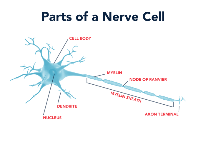
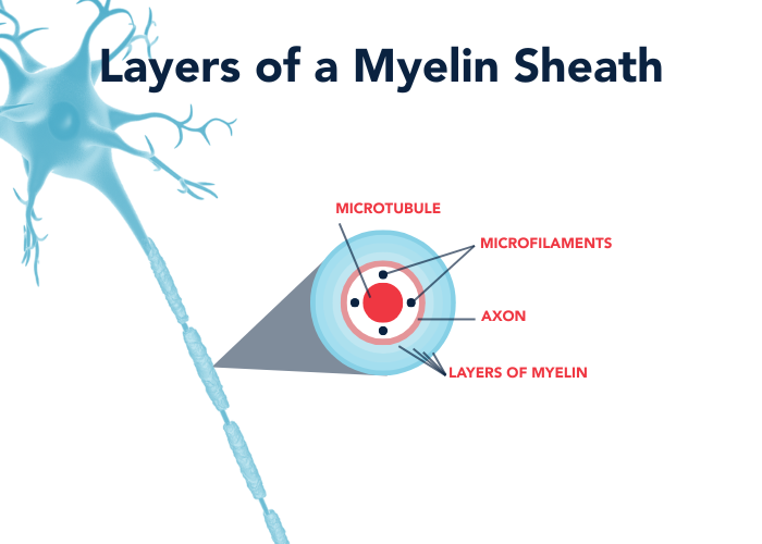
  • Neuronal changes: The structural shrinking and thinning is due in part to changes at the cellular level. Specifically, neurons themselves shrink and retract their dendrites, the branching structures that receive electrical signals. They also experience deterioration of myelin, the protective layer that coats their axons, which pass on signals. Neurons start to lose connections to other neurons across synapses. Finally, the process of neurogenesis, in which the brain creates new neurons to replenish cells that die, declines with age, amounting to a net loss of brain matter. 
  • Chemical changes: It appears that, as brains age, they generate and send fewer messenger chemicals like dopamine and serotonin. Though changes to dendrites also affect neurons’ ability to receive these neurotransmitters, lower production leads to reduced levels of these important chemicals, which can have effects on emotions and memory.

Brain Aging and Protein Protection

Another factor in brain aging can be seen at the molecular level. For proper internal functioning, healthy cells continually create, fold, dispatch, and recycle proteins, maintaining a balance called proteostasis. Research led by Stanford biologist Judith Frydman shows that when an aspect of this cycle slows down, it’s like a machine getting jammed somewhere in a factory production line; neurons end up with unfinished proteins that clump together and contribute to a slowdown in cognition. Every time there’s a disruption to our protein balance, it causes a loss of energy and potential damage that can accumulate over decades. 

Understanding this protein slowdown and its effects on aging could be helpful in the development of medications. Tests are underway for drugs like rapamycin, which targets the protein-making components of cells. It’s been successful at extending the lifespan of some mammals and shows promise in tests to establish potential biomarkers, or early indications, of Alzheimer’s disease. But if a drug could be targeted even more precisely to the assembly of proteins from amino acids, rather than the entire protein synthesis process, that could help patients avoid side effects like immunosuppression and difficulty in healing wounds. Theoretically, if medical science could keep the elongation process running at good pace, it could help prevent aspects of neurodegeneration. 

The Brain Age Gap and Predicting Alzheimer’s Progression

The difference between a person’s brain age and chronological age is called the brain age gap. A study by researchers in Tai’an, China, published this month in the Journal of Neurological Sciences, suggests that the brain age gap could be used to make early diagnoses of Alzheimer’s. This is because Alzheimer’s causes accelerated brain aging. If this widespread neurodegenerative disease could be detected early, it would be much easier to manage its progression. 

The researchers make the case that brain age can be estimated using neuroimaging and artificial intelligence (AI). With predictive models trained on healthy individuals, the researchers propose using “deep learning” AI to identify subtle changes in brain images that represent accelerated brain aging. Detecting a noticeable brain age gap could then become a useful biomarker of the early stages of Alzheimer’s. 

While this approach could be useful under the right conditions, the researchers acknowledge significant technical challenges that need to be overcome to make it reliable. They note issues like site bias, limited data, model accuracy, clinical applicability, and hardware demands. However, if these problems could be resolved through future research, it would mean that there would be a way to quantitatively identify brain aging consistent with Alzheimer’s, long before symptoms appear. 

Brain Aging and COVID-19

Last month, researchers at the University of Nottingham found that the COVID-19 pandemic may have led to a widespread acceleration in brain aging—even among those who never contracted the virus. The team reviewed longitudinal brain scans from nearly a thousand healthy adults, taken both before and after the pandemic. Using advanced imaging and machine learning, much like the researchers in the brain age gap study for Alzheimer’s above, the researchers compared brain scans to each person’s actual age. 

The changes that represented increased brain aging were most noticeable in older adults, in men, and in people from more disadvantaged backgrounds. The researchers discovered that only those who’d been infected showed signs of decreased cognitive abilities, but surprisingly, even people who never had COVID showed accelerated brain aging during this time. Dr. Ali-Reza Mohammadi-Nejad, the lead researcher, theorizes that the overall experience of the pandemic, including social isolation and constant uncertainty, may have influenced brain aging. This reinforces the idea that brains can age due to external environmental factors as well as internal biological ones. 

How to Slow Down Brain Aging

While brains can age faster than one’s chronological age, they can also age slower. It’s possible to be of advanced physical age and have a brain that’s healthier than average. For example, occasionally people in their eighties might have a brain age resembling what might normally be seen in people in their seventies. 

There are many reasons why the brain might age faster for some people, whether due to disease, genetic history, or environmental factors, but they are largely out of anyone’s control. With continued support for research, scientists will discover how to treat and eventually cure diseases that accelerate brain aging. In the meantime, there are things you can do to help prolong your brain health: 

  • Try to manage stress. Chronic stress has negative effects on many aspects of mental and physical health, including the immune and cardiovascular systems. It robs us of restorative rest, it leaves us feeling exhausted and unhappy, and it reduces our ability to focus and make decisions. Stress increases inflammation and cortisone levels in the body, which can cause damage to the hippocampus and increase the risk of neurodegenerative dementia. If it’s possible to change the life situations or environment that causes chronic stress, that would be very helpful for long-term brain health. If that isn’t possible, techniques to manage stress, such as mindfulness, meditation, regular exercise, and even deep breathing can make a positive difference.
  • Prioritize physical health. Get regular exercise, eat a balanced diet (ideally including omega-3 oils and antioxidants), and get enough good-quality sleep.
  • Practice cognitive activities and spend time with others. Engaging in activities like solving puzzles, learning new skills, and reading help to keep your brain’s cognitive abilities in good shape. To keep communication skills sharp, avoid isolation, and generally improve emotional health, it’s also important to maintain friendships and other social connections.
  • Get regular check-ups. Keeping up regular these healthy lifestyle habits will help your brain to be more resilient, but make sure to also see a doctor for preventative check-ups. Be open about how your body and mind are adjusting to life as you get older. Provide thorough information and complete the recommended health screenings. This data-driven approach to healthcare will help you be well informed to maximize your brain health in later years. 


The American Brain Foundation is committed to finding cures for all brain diseases and disorders. Donate today to make a difference. With your help, we can all experience life without brain disease.

Deep brain stimulation uses electrical pulses to affect signaling activity in specific areas of the brain. Initially developed in the 1980s, it’s most often used to treat Parkinson’s disease, essential tremor, and dystonia. The applications for this therapy have since extended to an increasing number of diverse neurological and psychological conditions. 


What is Deep Brain Stimulation?

Deep Brain Stimulation (DBS) is a surgical treatment that involves implanting electronic leads into specific parts of the brain and sending controlled electrical impulses to those regions. This stimulation modulates the electrical signaling that naturally occurs in the brain and central nervous system, and it can be used to either promote or disrupt those signals. DBS has proven to be a helpful therapy for some individuals with Parkinson’s disease, to assist with motor function and reduce tremor or other symptoms if medication is ineffective. However, not all patients with Parkinson’s qualify for DBS, and it’s generally not recommended for those who also experience dementia or severe depression. 

In some ways, DBS can be thought of as a pacemaker for the brain. Doctors first use preoperative advanced imaging techniques to determine which specific part of the brain needs electrical stimulation to alleviate the symptoms of a disease. A tiny opening is made in the skull, and a thin wire with an electrode at the end (called a lead) is threaded through to touch that precise brain area. The wire is implanted under the skin and connected to a small neurostimulator device implanted in the upper chest, typically near the collarbone. This device takes advantage of long-lasting lithium-ion battery technology, so that patients can retain their mobility. It automatically sends electrical pulses along the wire to the necessary brain area, when necessary, to stop activity associated with a brain disease symptom (such as a physical tremor) or excite neurons for proper electrical signaling. 

Deep Brain Stimulation for Parkinson’s Disease

In advanced stages of Parkinson’s disease, electrical signals in the brain become disorganized in areas that control movement, causing tremors and other motor issues. For many patients, medication is sufficient to control these symptoms. For those whose symptoms do not respond to medication, who experience side effects from medication, or who experience extreme symptoms as soon as the medication wears off, DBS might be a viable treatment. Electronic pulses sent from the neurostimulator interrupt these disorganized signals, stopping the motor symptoms of the disease.

DBS also has the potential to help Parkinson’s patients in a different way. Last year, a team of researchers based in Würzburg, Germany, and Toronto, Canada, conducted a study showing that DBS could be used to reverse the inflammation that drives Parkinson’s disease. Inflammation is a natural part of the body’s immune response, but sometimes this system malfunctions. Within the brain, inflammation can cause immune cells called microglia and astrocytes to mistakenly attack healthy brain cells, including those that produce the important neurotransmitter dopamine. In Parkinson’s, if enough of these dopamine-producing cells are lost, a person will experience motor symptoms like a tremor in their hands, though there may be other symptoms including loss of balance and coordination, sleep disturbances, sudden body movements or vocalizations, and mood changes. If this dysfunctional inflammation can be reversed through deep brain stimulation, it could provide relief and hope to many Parkinson’s patients. However, further study is needed to understand how DBS reduces inflammation. 

Deep Brain Stimulation for Epilepsy

Currently, medication is effective at controlling seizures for about 70 percent of people with epilepsy. For the remaining 30 percent, other types of treatment may need to be considered to stop seizures. DBS is one approach that has been studied to treat epilepsy alongside medication. 

Recently, the UK organization Epilepsy Action reported on trials of DBS that have been conducted in various countries to control epileptic seizures, particularly among patients with Lennox-Gastaut syndrome (LGS), which is a severe form of the disease. These trials have shown promise, and many of the participants ended up experiencing fewer seizures. A further study conducted at King’s College Hospital and Great Ormond Street Hospital in London observed the use of DBS to treat LGS in teenagers and children. The first pediatric patient to receive a DBS device to control LGS seizures experienced an 80 percent reduction in seizures. However, more work needs to be done to verify the efficacy and safety of this approach. Other pediatric patients are receiving DBS implants and devices in the United Kingdom over the next several years. 

Here in the United States, researchers at the Mayo Clinic have developed an advanced brain-monitoring system that uses artificial intelligence to continuously monitor brain activity in patients with drug-resistant temporal lobe epilepsy. This technology provides extremely accurate data to track and report seizures automatically (even during sleep) and administer DBS pulses when needed. The researchers also discovered that low-frequency DBS not only reduced seizures but also led to improvements in memory and sleep. 

Deep Brain Stimulation for Depression

In addition to its potential benefits for treating brain diseases that are traditionally categorized as neurological conditions, DBS and similar brain stimulation techniques such as Transcranial Magnetic Stimulation (TMS) show promise for treating disorders traditionally categorized as psychological conditions. These approaches have been studied as therapies for depression, post-traumatic stress disorder (PTSD), obsessive–compulsive disorder, and mood disorders. 

This year, at Mount Sinai West in New York City, a team of medical scientists became the first in the nation to launch a trial of a DBS implant to help patients with treatment-resistant depression. As the name implies, this is a severe form of depression that persists despite multiple treatment approaches, including psychological therapy, medication, and possibly other medical interventions. The use of deep brain stimulation to treat depression hasn’t been approved by the U.S. Food and Drug Administration yet, and its effectiveness and safety have yet to be established. However, the device used in this study was authorized for this type of clinical trial. The first patient who received this implant for depression will be observed for the next three years to see if DBS is effective in improving his symptoms. 

In some ways, DBS resembles a long-established technique, electroconvulsive therapy (ECT), which sends electricity into the brain. ECT deliberately triggers brief, controlled seizures for therapeutic effect, and it has proven successful for relieving the symptoms of Parkinson’s and treatment-resistant depression. ECT is often represented as a frightening procedure in movies, but in real life, patients are under anesthesia, the treatment is non-invasive, and it has been beneficial for many, many people. According to the National Alliance on Mental Illness, 70 to 90 percent of people who receive ECT for depression report an improvement after the treatment. Compared to ECT, DBS is still new, it requires invasive surgery to place wires in the brain and implant electronic devices, there’s a risk of complications from surgery (potentially including strokes), and it’s much more expensive. 


The American Brain Foundation is committed to finding cures for all brain diseases and disorders. Donate today to make a difference. With your help, we can all experience life without brain disease.

Strokes are emergencies that require immediate medical attention. While they sometimes have elusive causes, they’re often preceded by cardiovascular diseases or other conditions. Stroke risk also tends to be higher for those who experience a certain type of migraine or undergo one of several critical surgeries.


Strokes and Other Diseases

Brain health and heart health are directly related. In some ways, they’re inseparable. The brain requires blood to function, and if this blood supply is interrupted, the result is a stroke. If the interruption is caused by a blockage to a blood vessel, most commonly by the formation of a blood clot, this interruption triggers an ischemic stroke. If blood vessels in the brain swell, as in an aneurysm, and then rupture, this internal bleeding is called a hemorrhagic stroke or a brain hemorrhage. 

Heart disease, hypertension (consistently high blood pressure), atherosclerosis (the narrowing of arteries due to plaque buildup), and thrombosis (the development of clots in the bloodstream) are cardiovascular conditions that can directly lead to strokes. This is why cardiovascular disease and stroke share risk factors, such as diabetes, obesity, smoking, and high cholesterol. Genetic predisposition is also a common risk factor. 

Strokes can also be caused by other types of disease. Some infections and autoimmune disorders trigger inflammation that narrows arteries or causes blood clots to form. For example, rheumatoid arthritis, systemic lupus erythematosus (SLE), inflammatory bowel disease, psoriasis, and other inflammatory diseases have been associated with increased stroke risk, according to some studies. Additionally, strokes occur in about 6 percent of pulmonary tuberculosis cases and up to 20 percent of tuberculous meningitis (TBM) cases, in which the bacteria that causes tuberculosis infects the meninges, which are the protective membrane layers surrounding the brain. Strokes can potentially occur from bacterial infections like periodontitis, syphilis, and Lyme disease, and from certain viruses like chickenpox, HIV, and hepatitis C. 

Stroke and Migraine

An established link exists between strokes and migraine. These are two separate conditions, but they can have similar symptoms and share risk factors.  

While migraine can be a chronic condition, for most people the pain and sensory disturbances of migraine episodes are temporary. Strokes, on the other hand, can deprive brain regions of needed blood, which can cause cell death and permanent damage. Currently, there’s no evidence to suggest that a migraine can cause a stroke or vice versa. 

About 30 percent of people with migraine experience an “aura,” or symptoms lasting up to an hour that signal the beginning of a migraine episode, specifically visual changes, tingling feelings, vertigo, confusion, difficulty speaking or hearing, or momentary paralysis. Those who experience migraine with aura are about twice as likely to have ischemic stroke in their lifetime, compared to those who don’t experience migraine at all. The reason for this increased risk is not yet clear, but those who experience migraine auras should speak to their doctors about other ways to mitigate stroke risk, including addressing other potential health conditions like high blood pressure, diabetes, atrial fibrillation (a heart rhythm disorder), or high cholesterol. Women who experience migraine with aura are usually advised against using the combined oral contraceptive pill (containing both estrogen and progestin), because this can cause blood clots and increase their risk of stroke. In rare cases, strokes and migraine attacks can happen simultaneously. This event is known as migrainous infarction, and it tends to happen along with prolonged aura symptoms. 

Sometimes migraine auras can be confused with transient ischemic attack (TIA), a brief mini-stroke. For example, in migraine, headache pain is often on either the left or right side of the brain, which can also be the case with some kinds of stroke. However, migraine aura symptoms tend to develop gradually, while a TIA happens suddenly and often lasts for less than five minutes. If you experience a sudden severe headache or paralysis, especially on one side of the body, with no apparent cause, or other symptoms including slurred speech, dizziness, temporary blindness, confusion, it’s important to see a doctor right away. 

Unexplained Strokes

While most ischemic strokes are caused by a clot that forms and blocks the flow of blood to the brain, sometimes they can be idiopathic, or arise without a clear explanation. These are sometimes referred to as cryptogenic strokes. According to the American Stroke Association, about one in three ischemic strokes are cryptogenic. 

While the causes of cryptogenic strokes cannot be easily determined, that doesn’t mean they happen spontaneously for no reason. There are numerous possibilities for a cause other than a typical blood-clot blockage, including the following: 

  • Irregular heartbeat (atrial fibrillation) 
  • Heart structure problems 
  • Hardening of the arteries 
  • Blood clotting disorder (thrombophilia) 

Diagnostic testing and blood workups may help doctors narrow down the potential reason for an unexplained stroke. Sometimes after a routine evaluation, doctors might need to collaborate with other specialists, such as neurologists, cardiologists, and electrophysiologists to identify a definitive cause. However, following doctors’ advice for leading a healthy lifestyle should help reduce the risk of both standard and unexplained ischemic strokes, or reduce the chances of additional strokes in the future. 

Strokes after Surgery

When a stroke occurs during surgery or within 30 days after surgery, it’s referred to as a perioperative stroke. The overall incidence of such strokes is relatively low, about 1 percent in most types of surgery, and between 5 and 10 percent in cardiac, major vascular, and neurological procedures—though it can be up to 16 percent for the removal of glioma brain tumors. Perioperative strokes are serious complications that can be disabling or even deadly for patients. 

Statistically, perioperative strokes are increasing in prevalence, simply due to demographic shifts. Specifically, there’s currently a large population of aging patients with multiple chronic vascular conditions, who tend to be more eligible for high-risk surgeries. The most significant risk factors for this kind of stroke are as follows: advanced age, a prior history of stroke or TIA, atrial fibrillation, carotid stenosis (a narrowing of the carotid arteries), blood clots in the heart, or having two or more simultaneous cardiovascular issues. The risk of stroke depends on the type and complexity of the procedure. Perioperative strokes are mostly ischemic rather than hemorrhagic, and a potential complication that can lead to stroke is intraoperative hypotension, which is when blood pressure drops significantly during surgery. However, about 1.8 percent of “clipping” or “coiling” procedures to resolve unruptured brain aneurysms lead to hemorrhagic strokes.   

It’s important to keep in mind that surgeries with the potential to cause perioperative strokes are often necessary to save a patient’s life. Most doctors, patients, and their families would agree that this priority justifies the relatively minor risk of stroke. With continued research, advanced training, and future advances in technology, it’s likely that the risk of perioperative stroke will decrease. In the meantime, surgeons can best prevent perioperative strokes through comprehensive preoperative assessments and optimization, procedure-specific precautions during surgery, and systematic postoperative monitoring, so that potential strokes can be detected early and attended to promptly.


The American Brain Foundation is committed to finding cures for all brain diseases and disorders. Donate today to make a difference. With your help, we can all experience life without brain disease.

Nerve fibers are protected with a coating called myelin. Multiple sclerosis, other immune diseases, strokes, injuries, and inflammation from viral infections can damage this coating, and this process is known as demyelination. When myelin is worn down all the way, nerve fibers themselves can become damaged, leading to slowed or blocked messages between the brain and body.


What is Myelin?

Nerve cells throughout your body, including the brain and spinal cord, have three main components: a cell body, dendrites, and axons. Dendrites are rootlike structures branching off from the cell to receive and process electrical signals. An axon is the cell’s structure for transmitting signals to the next cell’s dendrites, and it resembles a long tail. Many axons are insulated with myelin, a fatty substance that protects the internal nerve fibers. It also helps with electrical conduction, so that messages to and from the brain can be delivered as quickly as possible. 

Diagram of a nerve cell, showing the nucleus at the center of the cell body with dendrites extending outward. The axon extends from the cell body, wrapped in a myelin sheath with gaps called nodes of Ranvier. Microfilaments run within the axon, ending at the axon terminal.
A diagram showing the anatomy of a nerve cell.

You might think of myelin as being like the insulated coating on electrical wires. However, myelin coatings feature gaps (called nodes of Ranvier) at regular intervals, which separate them into segments called myelin sheaths. This separation helps transmit signals faster due to a process called saltatory conduction, in which the signal “jumps” from node to node, at a speed of up to 150 meters per second. High-velocity signal transmission is important, because some individual nerve axons have remarkable lengths. They can be up to three feet long in humans and up to fifteen feet long in giraffes! 

Myelin is composed of membrane layers that are built from organic compounds called lipids, as well as proteins and water. In the brain and spinal cord, myelin is produced by separate cells called oligodendrocytes, which wrap around axons. The central nervous system relies on these cells to replace damaged myelin (a process called remyelination), but this method of repair tends to be slow compared to the rate of demyelination in brain diseases. 

When Myelin Wears Down

Brain diseases, neuroinflammation, strokes, and viral infection are some of the main causes of demyelination. The disorder most frequently associated with demyelination is multiple sclerosis (MS), a chronic, disabling condition that currently affects more than 2.9 million people worldwide and nearly 1 million in the United States, according to the National MS Society. Research indicates that MS involves an autoimmune response, in which the body’s immune cells mistakenly attack the myelin sheaths of neurons. This damages the myelin until the underlying nerve fibers are exposed, and then they are damaged as well.

Cross-section of a myelin sheath, showing concentric layers from the outside in: myelin layers surrounding the axon, which encases microfilaments, which in turn surround a central microtubule.
A cross-section of a myelin sheath.

When axon fibers are damaged, this slows down or blocks transmissions of electrical signals to and from the brain, leading to a variety of possible symptoms, which vary from person to person. These symptoms can be temporary or permanent, but they include the following: 

  • Fatigue 
  • Muscle weakness 
  • Numbness  
  • Vision changes (potentially even blindness) 
  • Cognitive changes 
  • Behavioral changes 
  • Difficulties with movement (potentially even paralysis) 

Demyelination can also occur from deficiencies of nutrients like vitamin B12 and copper, as well as electrolyte imbalances. Exposure to certain toxins can cause demyelination, as can vascular disease or a lack of oxygen to the brain, a process called hypoxic-ischemic demyelination. Spinal cord injuries can sometimes lead to demyelination as well. 

Brain Diseases Involving Demyelination

Any disease that causes damage to myelin is known as a demyelinating disease. There are multiple types of MS, such as classical MS, acute (Marberg-type) MS, neuromyelitis optica (Devic’s disease), and concentric sclerosis. These all involve demyelination in the central nervous system. 

Besides MS, demyelination also occurs as a part of other comparatively rare diseases: 

Inflammation from the immune response to bacteria or viruses:

      • Acute disseminated encephalomyelitis (ADEM)
      • Acute hemorrhagic leukoencephalitis (AHLE) 
      • Progressive multifocal leukoencephalopathy (PML) 

Inflammation of the spinal cord and/or optic nerve:

      • Transverse myelitis (TM)
      • Optic neuritis
      • Myelin oligodendrocyte glycoprotein antibody-associated disease (MOGAD)

Diseases in which the immune system attacks the peripheral nervous system:

Diseases in which the immune system attacks the pons region of the brain:

      • Central pontine and extrapontine myelinolysis (CPM and EPM)

Inherited diseases:

Demyelination Research

Researchers continue to study the mechanisms of demyelination of nerve cells. This focus helps increase expand scientific knowledge about how demyelination happens and what specific risk factors may be involved. Research like this could lead to potential preventative interventions. For example, a study late last year proved the theory that when the body fails to repair myelin, it leads directly to cell death. This is significant, but the project also identified a specific protein signaling pathway that was more active in animals that failed to repair myelin. Blocking that pathway prevented the loss of nerve cells.  

In addition, researchers have been making impressive progress in learning how demyelination might be reversed, potentially leading to new treatments or cures for demyelinating diseases. 

As mentioned in a recent American Brain Foundation blog, scientists are also researching ways to accelerate natural myelin repair. Researchers at the University of Colorado Anschutz identified an experimental drug that accelerates remyelination and has even restored lost sight in mice. 

At Cambridge University, researchers similarly working with mice found that by transplanting neural stem cells to the site of MS-like nerve damage, they could grow new myelin-producing oligodendrocytes. These cells were then able to form new myelin sheaths. 

The American Brain Foundation supports research into demyelinating diseases, and in 2022 it awarded a Next Generation Research Grant to Sachin Gadani, MD, PhD, who set out to determine why MS patients do not experience effective remyelination. 


The American Brain Foundation is committed to finding cures for all brain diseases and disorders. Donate today to make a difference. With your help, we can all experience life without brain disease.

Have you noticed that you tend to dwell on negative events and impressions more than positive ones? This characteristic of the human brain may have evolved to help us prioritize survival, but it can also influence emotion, causing stress, depression, and anxiety. Luckily, there are healthy ways to develop positive thinking for a more balanced perspective.


Why Are We Biased Toward Negativity?

Let’s say you receive both a compliment and a criticism the same day. Which are you more likely to focus on and remember? Most people would say they dwell on the criticism, even if it’s comparatively mild. This is because the human brain has a tendency to react more strongly to negative stimuli, an effect that has been recorded in brain scans using functional magnetic resonance imaging (fMRI). As result, negative experiences often feel more psychologically intense than positive ones, even if they carry equal emotional weight. This is known as negativity bias. 

In our evolution, negativity bias may have developed as a mechanism to protect us from harm and ensure our survival. For example, with brains that are highly attuned to recognize threats, we can more easily avoid those perils and continue our lives in safety. Certainly, this would have helped to protect early humans from predators and other dangers. But it may also have a contemporary role in early childhood development, as infants learn to recognize aversive stimuli. To a certain extent, negativity bias continues to help people recognize and reduce potential risks throughout their lives, playing a role in making careful decisions and improving safety precautions. 

If we find ourselves thinking a lot more about a negative aspect of an experience, we give that aspect more attention and are more likely to commit it to memory. A potential upside of negativity bias is that it can lead us to identify problems where we could make positive changes. Much like how we often learn best from our mistakes, focusing on our negative feelings can lead us to stop bad habits, address problems, and find solutions.  

But for many people, negativity bias can be so strong that, to them, nearly everything seems bad or dangerous. This can make life feel exhausting, difficult, and unhappy.

The Effects of Negativity Bias

Those who have very high negativity bias are prone to experience very high anxiety. Being in a state of heightened anxiety for extended periods of time can interfere with work, relationships, and daily life, and it can have a negative effect on mental and physical health. Chronic anxiety causes physical stress to the nervous, cardiovascular, digestive, immune, and respiratory system, and it has the potential to develop into a psychological anxiety disorder. It negatively affects concentration, mood, blood pressure, and breathing. It can also cause panic attacks, chest pain, rapid heartbeat, muscle aches, loss of libido, upset stomach or nausea, headaches, and depression. 

Anxiety triggers the release of hormones like cortisol and adrenaline, increasing your pulse and breath rate so your brain gets more oxygen. This is designed to help you act quickly in emergency situations. Normally, the effects of these hormones subside once you no longer feel anxious, but if anxiety remains constant, your body can’t return to normal functioning. Over time, this can weaken your immune response. 

High negativity bias doesn’t always lead to anxiety. For some, it can lead to isolation and depression, or difficulty sustaining friendships with others. And seeing life through a negative lens can distort our perceptions of reality, so that we end up ignoring its positive aspects. 

Because one of the evolutionary functions of negativity bias is to help reduce risk, excessive negativity can make people overly risk averse. This can lead to social isolation, loneliness, and feeling stuck. In this way, excess negativity can be an impediment to personal growth.

How to Be More Positive

The brain may have an evolutionary predisposition toward negative thinking, but it’s still possible to manage negativity bias and reduce the effects it has on our mood and well-being. This can be accomplished by developing stronger positive thinking habits and considering alternate perspectives. These approaches don’t diminish your ability to recognize and respond to danger, nor do they encourage ignoring the effect of truly negative experiences. They’re simply methods that could be effective in helping to bring more emotional balance to your life.  

In a recent article for The Washington Post, writer Katherine Kam interviewed experts on negativity bias, who provided the following recommendations for finding this balance: 

  1. Shift your attention. When we have negative thoughts or experiences, sometimes it can be helpful to avoid generalizing and instead look at another aspect that’s positive. Negativity bias can blind us to the positive things happening around us all the time, so it helps to practice noticing them. 
  2. Make a habit of gratitude. One way to train your mind to be more positive is by setting aside a bit of time each day to write down some things you’re grateful for. You might find this difficult at first, but over time it gets easier, and then it can become automatic, and you’ll be more attuned to recognize positive experiences in the moment. 
  3. Harness negativity to break bad habits. If you have negative feelings about your own unhealthy habits—such as smoking, drinking too much alcohol, or eating too much sugar—focusing on those feelings can help strengthen your motivation to make positive behavioral changes. 
  4. Turn negative thoughts into positive action. Ruminating on negative experiences is very self-focused, but a productive way out that rut is to redirect your attention to connecting with and helping others who might be experiencing similar problems. 

Psychologist Catherine Moore suggests some additional strategies: 

  1. Pay attention to your thoughts and challenge negative self-talk. When you have a negative thought, whether it’s about a situation, another person, or yourself, try to identify if it helps you in some way or merely causes you anger, resentment, or frustration. Consider if your negative thought might be informed by an unfair judgment, jumping too quickly to a conclusion, or negativity bias. 
  2. Practice mindfulness. Through quiet reflection, or listening to guided meditations, you can become more attuned to your own emotions and how you experience them. When you have negative thoughts, take the time to breathe slowly and deeply. Observe and think about your feelings. 
  3. Reframe the experience. Psychologists often use a technique called cognitive behavioral therapy (CBT) to help patients cope with negative thoughts and beliefs. This involves a process of restructuring thoughts, “reframing” them from a more objective neutral perspective, and trying to withhold initial judgment. 
  4. Savor positive moments. When you have a positive experience, take the time to really appreciate it and how you feel. Take note of what you enjoyed about the experience and reflect upon it later. Getting into the habit of savoring such moments helps you remember them, and recalling positive thoughts can help provide balance when negative thoughts arise. 


The American Brain Foundation is committed to finding cures for all brain diseases and disorders. Donate today to make a difference. With your help, we can all experience life without brain disease.

Elizabeth DeStefano has lived with migraine for almost her entire life. In searching for help, she came across the Migraine World Summit, an annual conference for people with this condition. She became an active volunteer with the organization and this year co-hosted the event. As a passionate advocate, she emphasizes the positive ripple effect of investing in research to improve life for those affected by migraine.


Elizabeth’s Experiences with Migraine

Elizabeth DeStefano has lived with migraine for almost her entire life. She first received an official diagnosis when she was seven or eight years old, and she has continued to struggle with migraine attacks on a regular basis. 

Most people associate migraine with severe headaches, and that’s certainly a significant part of the experience for many, but there are other symptoms as well. “My migraine has changed a lot over the years,” Elizabeth says. “When I was younger, the symptoms that would accompany the often very severe head pain were nausea and vomiting and disorientation. I remember a time even in middle school, walking down the hallway … I was nauseous and experiencing what’s referred to as Alice in Wonderland Syndrome, where things are moving, things were sliding down the walls and changing in proportion and size. That’s not been a frequent occurrence for me, but something that I remember to this day from a middle school phase of life.”  

As she grew into adulthood, the symptoms that accompanied her headaches transitioned from nausea to other difficulties. These included increased sensitivity to light and sound and “sometimes [having] trouble finding a word, pulling that word down and grasping and not getting it right away. Or just a little lapse or a little bit of what you hear people talk about as brain fog, or any type of cognitive impairment where I just feel a little bit slower in speaking or formulating my thoughts.” She also noticed mood changes, such as increased irritability, or sudden fatigue, particularly during the prodrome, the phase of a migraine attack before the onset of pain. 

Not everyone who has migraine attacks experiences headache pain, Elizabeth explains. “But for most people, migraine occurs in three or four stages: prodrome, the pain phase, and then post-drome, which you sometimes hear discussed as the hangover phase.” The prodrome phase includes a variety of potential symptoms from neck pain to dizziness, fatigue, and difficulty concentrating, but it reflects the reality that migraine episodes begin well before headaches start. In the post-drome phase, the headache pain recedes, but there are often other lingering symptoms. For a day or more after she experiences a particularly bad attack, Elizabeth sometimes feels as if she has the flu or is recovering from a physical accident. Up to a third of people who experience migraine also experience a fourth phase called the aura phase, which occurs before the prodrome and serves as a warning sign of an impending episode. This phase often involves visual cues, such as the appearance of flashing lights or zigzag lines, blurry vision, or temporary vision loss, or it might include tingling sensations on one side of the body or an inability to speak clearly.  

A newer symptom Elizabeth has come to recognize for herself is ear pain on one side, which for her is an early warning that an attack is in progress. This reflects the reality that migraine is complex, often involving multiple symptoms that change over many years, and they are also unique for each individual. “As these symptoms change,” she says, “it can lead you down some rabbit holes as you explore things that you are sure must be other conditions, but that are actually part of your ever-evolving migraine disease.” 

Migraine World Summit Co-host Elizabeth DeStefano shares her personal experience living with migraine and explains why advocacy and research funding are essential to changing lives.

Living with Migraine

For many, intense migraine attacks can be severely debilitating, making it impossible for them to get out of bed or do much else beyond trying to endure the overwhelming pain. The fact that an episode like this could occur at any time leads to other problems, which are often overlooked. 

“It’s part of the burden of the disease, living with something that’s unpredictable and incapacitating potentially,” Elizabeth explains. The attacks are not only unpredictable in their timing, but also in their severity. An episode might make a person unavailable to spend time with others as planned, potentially causing feelings of guilt. Not knowing when a bad migraine episode might occur can lead to social withdrawal and depression. 

“There’s the emotional toll it takes too, which is, for me, this feeling through different phases of my life of feeling less than—less than the friend I want to be, less than the mom I want to be, less than the wife I want to be, less than the professional I want to be,” she says. “It’s that idea of not being able to live up to your potential, either professionally or socially or personally or all of those things.” 

People who experience migraine are often advised to focus on maximizing their sleep, nutrition, exercise, hydration, and stress management. These five lifestyle best practices are helpful for general health and well-being, but they’re usually insufficient to prevent or treat migraine episodes. For some, cognitive behavioral therapy can help with migraine management. Others may benefit from meditation and mindfulness, taking a walk in nature, or neuromodulation (using a specific device that sends electric stimulation to the brain to interfere with the signaling thought to be a part of migraine). There are also medications that can help prevent migraine episodes or reduce the severity of symptoms in the moment, but they’re not equally effective for everyone. Because an individual’s migraine experience is so unique, trial and error are usually part of the process of finding a healthy way to manage symptoms. But exercising caution is important. “When you’re desperate, and when you live with unpredictable and potentially incapacitating pain or disability, you can be easy prey for the latest thing that shows up on TikTok, or is promoted marketing,” Elizabeth says. Instead, she and others in migraine advocacy emphasize seeking out verified, evidence-based information. 

The Migraine World Summit

For a while, Elizabeth’s episodic migraine became chronic, with little if any recovery time between attacks. “It is a shocking thing. It should be a shocking thing that there’s a chance that you could become chronic from episodic migraine,” she says. “I had never been told about that. I had never had conversations with any of my providers about the fact that that was a possibility.”  

But then, in 2017, Elizabeth discovered the Migraine World Summit, an annual event that presents expertise on migraine free of charge. 

“I tried to make space for every possible minute of that eight days of the summit that I could, because I was absolutely astonished that first of all, this was offered to people, and for free no less,” she says. The summit offers eight days of interviews with world-leading experts in migraine, providing information that can be otherwise difficult for patients to access, especially considering that despite the prevalence of migraine in the United States, there are relatively few doctors who specialize in treating the condition. “I learned so much that first year,” she says. “I also was really touched that there were people who were making this possible. … I was so moved that they cared enough to make that possible for me and other people watching. I felt seen and heard and supported in a way that I hadn’t throughout my life.” 

After that year’s summit, a call went out for volunteers, and Elizabeth wrote back immediately, saying she’d love to lend any skills she had to this work. She started that same year, helping with the content-editing team, and she has progressed through various roles with the event since then. This year, she proudly served as the summit’s co-host. 

“What I’ve learned is that there is a world of migraine advocacy,” she says. “I didn’t know that there was this group of individuals, both clinicians, researchers, and people who live with migraine themselves, who work together in overlapping ways and in different areas to bring awareness and education of migraine and other headache disorders to the world. And I didn’t expect then in 2017 that I would become a part of that group of people, but I feel very privileged that I have since that time.” 

Through the summit, Elizabeth learned more about how episodic migraine can transition to chronic migraine, and how that change can be prevented and sometimes reversed. With a great deal of effort, she was able to transition back to episodic migraine. This involved a combination of medications, nonpharmacologic therapies, and lifestyle changes. It took time, self-education, and a lot of trial and error. 

Elizabeth is forever grateful that she had people in different phases of her life who understood and had compassion for what she was going through, but she also knows that not everyone is so fortunate. There are many people who don’t understand what it’s like to live with migraine, who often assume it’s just a standard headache, for example—or worse, simply an excuse to get out of a commitment. These misunderstandings underscore the need for wider education and advocacy, which is something that Elizabeth has taken to heart as a spokesperson. 

Progress is Possible Through Research

“Migraine is one of the most disabling conditions globally,” Elizabeth says. While it affects people of all ages, including children and older adults, it seems to disproportionately affect people at a stage in life when many other big things are happening or could be happening, such as raising families or advancing in careers. 

While migraine is disabling, it’s by no means rare. In fact, it’s the third most common disease in the world, affecting up to 12 percent of the global population. Despite this prevalence, funding for migraine research has been relatively scarce. “When you look at how much funding migraine and headache disorders receives compared to other disorders that affect far fewer people, it becomes very clear how underserved migraine research is,” she says. She believes that this is due in part to the fact that, historically, migraine severity has been greatly underestimated. In addition, some individuals who live with migraine may feel that there’s a stigma against advocating for themselves in a professional environment, especially when supervisors or others in the workplace might not have the context to understand the disease. 

However, Elizabeth acknowledges that even if research on migraine has been limited compared to other diseases, the work that’s been done has still made a significant difference. “There has been incredible research done in recent years in migraine that has given hope to the community of providers and researchers and people living with migraine that other treatments are possible,” she says. “With the advent of recent targets like CGRP,¹ there have been advances from a pharmacologic standpoint that have been absolute game changers” for many patients. 

Further progress is possible through continued research. “We know there are other targets that have not been identified,” she says. “And then we know there are other options that have shown to be effective that are outside of pharmacology that alone or maybe in combination with the pharmacologic option could be complete answers for people. So if we can look at this and realize that by giving people options through the research that is desperately needed, and the funding for that research that is desperately needed, we can make huge strides in this.” 

Elizabeth likes to emphasize ripple effects. Migraine affects individuals, but it also affects their families, social circles, colleagues, professional opportunities, health insurance, and so on. “The financial, social, and economic burden is profound. And if we could solve that even a little bit more, the ripple effects would be equally positive,” she says. And because research in one area of brain disease often leads to new insights about others, new discoveries about migraine could lead to improved outcomes for other disorders. 

“If we could figure this out, it would, without a doubt, bear fruit for figuring out so many other things, neurologic conditions and potentially beyond,” she says. 


The American Brain Foundation is committed to finding cures for all brain diseases and disorders. Donate today to make a difference. With your help, we can all experience life without brain disease.

A traumatic brain injury (TBI) occurs when a person experiences sudden external blunt force trauma, generally from a significant impact to the head or from violent jolts. Common causes of TBI are falls, motor vehicle crashes, firearm-related incidents, physical assaults, and sports injuries, and the severity can vary from a temporary concussion to permanent brain damage. Fortunately, research continues to explore treatments to help people recover from TBI. 


Traditionally, options for treating traumatic brain injury have been limited. In some cases, TBI may require brain surgery to repair hematomas (ruptured blood vessels) or contusions (bruised brain tissue). Even when it isn’t possible to heal physical brain damage from TBI, rehabilitation can often help patients regain much of their brain function, skills, and independence. Rehabilitation programs may include physical therapy, speech and language therapy, occupational therapy, and caregiver training. This often amounts to a long and involved process of recovery involving progression through several levels of treatment, beginning with acute care in a hospital setting, transitioning to rehabilitation in an inpatient facility, and eventually moving to in-home therapy services or the equivalent. 

All of this remains important in post-TBI care. However, research is leading to new insights that could significantly improve treatment after a brain injury. The following research studies could provide relevant details that might lead to better procedures for TBI recovery. 

Astrocytes Play Important Roles in Brain Repair 

The brain is made up of different types of cells, most of which are grouped into two categories: neurons and glia. The function of neurons is to communicate with other neurons, by sending signals throughout the brain and the entire nervous system. Glia, on the other hand, perform a variety of functions to support neurons. Although the adult human brain has an estimated 100 billion neurons, glial cells far outnumber neurons. One important function of the star-shaped glial cells called astrocytes is repairing the brain and spinal cord after an infection or injury. 

A study published in the journal Cells explores the role of astrocytes in further detail and suggests that there may be ways to medically enhance their ability to heal tissue after a TBI. The researchers, primarily based at the University of Lublin in Poland, explain how a TBI transforms astrocytes to put them into a reactive state, which enables them to perform beneficial tasks like stabilizing the blood–brain barrier, facilitating the production of neurons (neurogenesis), secreting helpful biochemical compounds, and lowering inflammation. However, this response to injury also has potential adverse effects. Astrocytes create scar tissue as a protective barrier between damaged and healthy tissue. That’s important, but this scar tissue has the downside of impeding cell regeneration and increasing inflammation. 

The researchers theorize that it might be possible to improve recovery from TBI by manipulating the mechanisms of human astrocyte behavior. In findings from animal model studies, it appears that naturally produced molecules, including endocannabinoids and a form of estrogen, show promise in terms of potentially enabling doctors to influence astrocytes’ regenerative functions. This could make it possible to boost the positive effects of the cellular response to TBI while reducing the negative effects. 

Wrapping Stem Cells with Wireless Electrical Nanopatches

The journal Nature Communications reports on a very different approach to improving TBI recovery: using implanted stem cells and nanotechnology. 

Imagine extracting neural stem cells from elsewhere in the brain, transplanting them to the area surrounding a brain injury, and encouraging them to form into healthy neurons. That approach might have seemed far-fetched not so long ago, but now it’s the focus of research. One immediate problem that would need to be solved for this to work is that the stem cells must differentiate themselves correctly as they grow, forming neurons of a particular type that serve particular functions. 

Electrical stimulation can enhance the process of differentiation for neural stem cells so that they can heal damaged brain tissue. But unfortunately, stem cells lose electrical stimulation after they’re transplanted. So a team of researchers primarily based at Shandong University in China tested a novel idea. They theorized that if the stem cells could be “wrapped” with microscopic electrical “nanopatches” made of conductive graphene nanosheets, it might be possible to use electromagnetic induction to send wireless electrical stimulation to those patches and control the growth of those stem cells. In this way, the stem cells could differentiate into the intended type of neuron to treat TBI—or possibly other types of brain diseases that result from neurodegeneration. 

To test this idea, the scientists worked with mice that had brain injuries. Those mice received implanted stem cells with and without nanopatch wrapping, and the ones with patched cells were exposed to a rotating magnetic field for 30 minutes a day. Within five days, 41 percent of the wrapped stem cells differentiated themselves into functional neurons, as opposed to only 16.3 percent of the unwrapped stem cells. The study reports that compared to the mice with unwrapped stem cells, the mice with electromagnetically stimulated wrapped stem cells showed significant recovery of brain tissues, behaviors, and cognition within 28 days. 

Traumatic Brain Injury Could Be a Risk Factor for Parkinson’s 

A study published in Molecular Neurobiology this year explores the links between TBI and the development of Parkinson’s disease (PD). Back in 1817, when James Parkinson initially described the condition that now bears his name, he hypothesized that head trauma could be a cause. However, debate has continued ever since about the relationship between TBI and PD, and there haven’t been many research projects to determine what would be involved if TBI can lead to PD.  

The study here, from three researchers at the University of Adelaide in Australia, goes into detail about shared characteristics between TBI and PD, and indications that TBI correlates with an increased risk of developing PD. The study considers two neurotransmitters (signaling molecules) that travel across synapses (spaces between neurons), specifically dopamine and noradrenaline. It also highlights a protein called α-synuclein.  

One of the most notable characteristics of PD is a loss of neurons in a part of the brain called the substantia nigra. These neurons produce and release dopamine, which is involved in controlling the body’s movements. This is why people with advanced PD often have motor problems such as tremors, muscle stiffness, and difficulty walking. Preclinical evidence shows that TBI leads to similar neuronal loss and other dysfunction related to dopamine.  

Secondly, the neurotransmitter noradrenaline, also known as norepinephrine, decreases in PD. Noradrenaline is critical for managing alertness, responding to stress, controlling blood flow when needed, storing and retrieving memories, and operating the body’s fight-or-flight response in dangerous situations. The part of the brain primarily responsible for producing noradrenaline is the locus coeruleus, and this cluster of neurons is notably disrupted in patients with PD. Research suggests that after a TBI, the locus coeruleus initially produces excess noradrenaline to help protect the brain as it tries to heal, but then the noradrenaline in this region decreases to baseline levels or falls below baseline.  

Thirdly, both PD and TBI involve an accumulation of α-synuclein proteins. These large molecules are primarily responsible for repairing DNA, but they can sometimes become “misfolded,” clump together, and form structures called Lewy bodies within neurons. These structures can continue to grow with the continued accumulation of misfolded α-synuclein, to the point where they eventually destroy the cells they accumulate in. This loss of cells causes the symptoms in Parkinson’s, Lewy body dementia, and some other neurodegenerative diseases. 

It’s important to note that traumatic brain injury doesn’t always lead to Parkinson’s disease, and Parkinson’s has a variety of causes, including genetics and heredity, lifestyle, and environmental factors (a topic studied by American Brain Foundation Next Generation Research Grant recipient Brittany Krzyzanowski, PhD). Nevertheless, practicing appropriate safety measures—such as wearing a helmet when playing contact sports or riding a horse, motorcycle, or bicycle—might help to reduce the risk of developing Parkinson’s later in life. 


The American Brain Foundation is committed to finding cures for all brain diseases and disorders. Donate today to make a difference. With your help, we can all experience life without brain disease.

In May, the Eyal Kimchi Lab at Northwestern University’s Feinberg School of Medicine in Chicago graciously welcomed American Brain Foundation staff and board members to see translational neuroscience research in person. Dr. Kimchi is one of the Foundation’s past grant recipients, and he has gone on to lead a team of researchers who are working to better understand and treat delirium. Visiting the lab was an excellent opportunity to learn about scientific methods and experiments and to hear more about how grant funding can kickstart a fulfilling career in research.


The Value of Early-Career Research Grants

Dr. Eyal Kimchi received an American Brain Foundation Next Generation Research Grant in 2014. Last month, after welcoming the staff and board members to Northwestern, he began the program for the afternoon site visit with a clear and inspiring presentation. His talk covered the impact that this grant had on the trajectory of his own career. 

“I became very interested during medical school in this condition called delirium,” he explains. Delirium is defined as a state of acute confusion with disturbances in attention, awareness, and cognition. He found it striking that it can present so dramatically with patients, yet there were no approved therapies to treat it. “It is a dramatic change in our brain and mental function, and we don’t have a place to take someone to other than the hospital where at that point, as doctors, we’re a little bit lost. So that has really motivated a lot of my career.” 

Dr. Kimchi continued with his studies and learned that one reason why delirium has been under-researched is that it can be difficult to categorize. “Delirium is a disorder that falls between a lot of different specialties,” he says. But he knew that it was a topic that needed to be investigated. After completing his degree, he applied for an ABF early-career research grant, with a proposed study that didn’t fit neatly into a pre-defined disease classification. “What really had a tremendous impact on me was that the American Brain Foundation didn’t care about that. They didn’t care about what funding agency it fell into, or what specialty it fell into. They agreed with the proposal that this was an important brain problem, and it needed a diversity of approaches to be applied to it.” 

Even if his proposal was a bit of a “square peg,” as Dr. Kimchi puts it, the selection committee recognized the significant impact the study could have for patients in the future. They awarded him the funding he needed to set aside the time to develop a feasible research model using rodents. The funding enabled him to figure out how to induce and measure delirium in mice and rats. This research data made it possible to secure further funding from the National Institutes of Health (NIH) for hypothesis testing. He also credits the American Academy of Neurology’s career-development workshops for providing valuable advice on how to build on research to obtain continued funding. 

The visit highlighted the multigenerational continuum of researchers involved in this work. Dr. Merit Cudkowicz was the Foundation’s very first Next Generation Research Grant recipient, and she has moved on to become not only one of the world’s foremost researchers and experts on amyotrophic lateral sclerosis (ALS), but also a board member with the Foundation. In between, she taught Dr. Kimchi when he was a medical student and championed his innovative work. Dr. Cudkowicz was part of the visiting group, and the two doctors greeted each other warmly. This mutual respect also extended to the brilliant and dedicated post-doctoral, graduate, and undergraduate researchers who work at the lab. 

What Is Delirium?

Delirium is a condition that results in confused thinking and lack of awareness of one’s surroundings. It can affect a person’s memory, speech, and comprehension, and it can cause behavioral and emotional changes, such as anxiety, fear, distrust, depression, anger, elation, restlessness, sluggishness, sudden mood swings, changes in sleep habits, and sometimes even hallucinations. The symptoms can be difficult to distinguish from those of dementia, but delirium typically has a comparatively quick onset (between a few hours and a few days), and the effect is usually temporary. Additionally, it impairs a person’s ability to maintain focus on a given thought or topic, whereas in the early stages of dementia, people can usually pay attention without being sluggish or agitated. Delirium symptoms also change rapidly and come and go throughout the day. Dementia, on the other hand, represents a more gradual and consistent decline due to neurodegeneration. Both are common disorders, and a person may have both at once. 

Delirium has many causes. It can occur as a side effect of a severe or long-lasting illness; it can result from a chemical imbalance in the body, such as low sodium; or it can be caused as a side effect of medications, infections, surgery, or alcohol or drug use or withdrawal. Notably, it is common among hospital patients, and according to the Kimchi Lab team, about 22% of all hospital patients have bouts of delirium, and it is especially prevalent among intensive-care unit (ICU) patients. Statistically, delirium doubles the risk of mortality, and the experience can be disorienting and frightening for patients and their loved ones. Even so, the condition has been understudied.  

The Kimchi Lab team has narrowed its focus to two major types of delirium, one related to systemic inflammation from the immune system, and the other related to chemicals in the brain called neuromodulators, specifically acetylcholine. They are working to understand what’s really happening within the cells and neural network when a patient becomes delirious. 

Four Stations of the Lab

Following Dr. Kimchi’s presentation, ABF staff and board split up into four groups to take turns visiting with members of the research team who specialized in one of four areas: 

  • Mouse brain activity
  • Mouse behavior
  • Wearable wireless EEG devices
  • AI analysis

The lab has refined Dr. Kimchi’s model for observing the effects that delirium has on rodents. Now specifically using mice, one group of researchers monitors a mouse with induced delirium walk on the exterior surface of a specially designed exercise wheel, which it uses at its own pace. The mouse is gently kept in place by an exterior structure, and wires connect tiny electrodes to its brain. A small camera is set up near the mouse’s head. With the electrodes and camera, the team can see not only its brainwaves, but also which areas of the brain show electrical activity at any given moment. 

Separately, another station monitors the behavior of 40 mice in 24/7 live video feeds. Each mouse is situated in its own identical container, and the researchers compare the behavior of delirious and non-delirious mice. Since it would be impractical for humans to watch all this video footage to collect data, the team employs computer systems with motion-capture technology to monitor various coordinates on a digital overlay of each mouse’s body. Then these movements are recorded and analyzed using artificial intelligence (AI). In this way, all the mice can be monitored for the entire duration of testing. 

For both mice experiments, the team follows strict ethical standards to ensure that the animals are treated with the utmost care. 

The other two stations are working on translational applications for humans with delirium. One team is focused on refining and using a new kind of wireless electroencephalogram (EEG) device for patients with delirium. Traditional EEG devices have more than twenty electrodes for monitoring the scalp for brain activity in different regions, but they are cumbersome for a patient suffering from delirium. So the team developed a smaller, battery-powered, wireless EEG with only two electrodes, which sits comfortably on top and toward the back of a patient’s head. The impressive device was demonstrated in real-time, showing a researcher’s brainwaves appearing on a tablet. 

The fourth station focused on computer analysis of data from human patients. One issue with diagnosing delirium the old-fashioned way—by asking the patient questions—is that it’s subject to inherent inequity and bias. For example, patients may not always speak the same language as doctors. The researchers’ aim is to develop novel, highly sensitive, and general tools to evaluate delirium that are equitable and free of bias, and their approach is to use video-EEG monitoring. This combines video of patients and simultaneous EEG monitoring. With this data, the team can analyze a patient’s movements and posture in detail. AI is then used to try to predict movements that can be associated with delirium. The researchers in these roles also analyzed EEG brainwave activity from patients with delirium by converting the waveform results into colorful two-dimensional representations called spectrograms. In this configuration, it was clear that delirium produces slow brainwave activity resembling sleep, and that sleep-wake patterns of brainwave activity are affected. 

Funding Makes Research Happen

With recent cuts to the NIH budget for funding biomedical research, important studies like those at the Eyal Kimchi Lab are under threat. In his presentation, Dr. Kimchi spoke of other programs that have tragically lost their funding to continue their work. The lab is currently still going strong, but at the time of the visit, Northwestern hadn’t received a single dollar from the NIH since mid-March for grants that were already awarded (and not cancelled). This budget-cutting has global consequences, and scientists worldwide are concerned about the costs of halting progress in U.S.-funded brain disease research.  

Is there hope? “For better or worse, we are being forced broadly as a research community to [better] communicate the importance and value of what we do,” Dr. Kimchi says. “One thing that gives me hope is seeing people understanding and pivoting toward the importance of this broad-based advocacy.” Besides researchers becoming more engaged in advocacy, he sees advocacy organizations becoming more savvy about the infrastructure and business of research funding in the United States. “We’ve been kind of siloed. ‘I’ll just do my research, you’ll just do the advocacy for me, and we’ll meet over the dollars.’ And I think that we’re all getting a better sense of how to integrate shared priorities.” 

In the meantime, he encourages researchers to keep applying for grants, including those from the American Brain Foundation. The researchers who receive grants are all scientists who believed in their proposals and took the effort to apply. But Dr. Kimchi points out that there are benefits to applying beyond potentially receiving funding. “I was so appreciative of the feedback I got on my application,” he says. “The feedback I got was still so helpful for the next grant. And I have a mentee in my lab right now who applied to an ABF fellowship last year, and didn’t get it, but the feedback he got was so helpful and has really shaped [a different] application he just submitted and the [ABF] application he’s going to submit again this year. It is a real opportunity to have your thoughts and ideas and dreams be reviewed critically and constructively by world leaders [in brain research]. … It’s a smaller gift, but it is not a trivial gift. It is an important gift.” 

The American Brain Foundation is extraordinarily grateful for Dr. Kimchi and his colleagues for their hard work, as well as their kindness and hospitality. The ABF staff and board were also touched to feel so appreciated and welcome. “I am unbelievably appreciative to the ABF,” Dr. Kimchi says. “It served me at a time when I felt vulnerable and uncertain. I can see it continuing to play so many important roles.” 


The American Brain Foundation is committed to finding cures for all brain diseases and disorders. Donate today to make a difference. With your help, we can all experience life without brain disease.

Huntington’s disease is a progressive neurodegenerative disorder caused by an inherited mutation of a specific gene. Scientists now have a new understanding of how this mutation leads otherwise healthy individuals to develop symptoms later in life. 


What is Huntington’s Disease?

Huntington’s disease (HD) is a rare genetic disorder that affects about 1 in 10,000 people in the United States, and parents with this condition have a 50 percent chance of passing it on to their children. It is caused by a mutation of the HTT gene, which produces a protein called huntingtin. Our entire genetic code is made up of combinations of four building blocks called nucleotides, which are named adenine, cytosine, guanine, and thymine, or A, C, G, and T for short. The genetic code features mostly three-letter sequences of these nucleotides, one after the other, and one of the combinations that features in HTT is CAG. These repetitions of CAG create elongated versions of huntingtin, which then accumulate in brain cells and ultimately kill them, leading to spreading neurodegeneration. 

Text: Nucleotides within DNA. Image: A magnified image of a section of a DNA helix showing how combinations of adenine, cytosine, guanine, and thymine make up the spokes between the outside of the helix.
A diagram showing how our entire genetic code is made up of four nucleotides called adenine, cytosine, guanine, and thymine. These are called base pairs when referring to a combination of two nucleotides that are joined together.

Most people inherit the HTT gene with 15 to 35 consecutive CAGs and never develop HD, but those who inherit a version with 40 or more consecutive CAGs will almost certainly develop the disease later in life, between the ages of 30 and 50. The more consecutive CAG repeats there are in DNA, the younger a person tends to be when symptoms start to appear. The symptoms include a variety of movement, cognitive, and psychological issues. HD can cause uncontrolled twitching, jerking, and writhing movements of the arms, legs, face, head, and torso, collectively known as Huntington’s chorea. Impairment of eye movement, balance, walking, speech, and swallowing are common. Mental processing and emotional regulation are also negatively affected. 

HD is a neurodegenerative disorder, and as more and more brain cells are destroyed by excess huntingtin protein, the symptoms worsen, and the disease becomes fatal. Currently, treatments exist to manage the symptoms, but a means to stop or reverse HD progression has yet to be discovered. 

A New Understanding of Huntington’s Disease

This past January, a study published in the journal Cell provided new insight about HD. Scientists at Harvard Medical School, the Broad Institute of MIT and Harvard, and McLean Hospital developed a new technology for analyzing gene expression in thousands of cells at once. They then expanded upon this technology to measure the length of the repeating series of CAG in individual cells.  

What they discovered was that these long repeating sequences aren’t inherently harmful, but they are unstable. As a person ages, the series of repeating CAGs gets longer and longer, and it only begins to cause harm when neurons have genes with more than 150 repeats. However, by using computer modeling of the collected data, the team was able to figure out that the rate of the expansion in the number of repeats begins to accelerate rapidly at a certain point. It takes a few decades for a person to go from 40 to 80 CAG repeats in their HTT genes, but once the number gets above 80, it begins expanding faster, and in only a few more years, the number nearly doubles and leads to cell death. This sudden acceleration provides an explanation for why HD symptoms appear only later in life. 

Beyond enhancing scientific understanding of how mutations lead to diseases, this study suggests future opportunities to study possible ways to treat HD. The authors note that the drugs that have been developed so far to reduce activity of the mutated HTT gene have struggled in clinical trials, and the reason may be that only a small number of cells have the mutated version of the gene or its produced proteins at a given time, so the treatment is helping only a small number of cells. But if a method could be devised to stop or slow the expansion of CAG repeats in the gene, the authors believe it might be possible to delay or even prevent the onset of the disease. 

Could a Common Heart Drug Help?

Another study, by a team at the University of Iowa, have reported that beta-blocker drugs, which are commonly used to treat heart and blood pressure issues, showed success in slowing the onset of HD symptoms. Their research, published in the journal JAMA Neurology, suggest that norepinephrine, the neurotransmitter and hormone involved in the human “fight or flight” response, may play a role in HD. The team’s previous research had found that patients with HD tended to have a stronger “fight or flight” reflex than people without the disease, even while at rest. 

They hypothesized that because of this stronger reflex, and by extension, a slightly more active sympathetic nervous system, HD patients might have elevated levels of norepinephrine. If so, a drug that inhibits that neurotransmitter might be therapeutic for those individuals. To investigate the possible benefits of beta-blockers, the team combed through data from the world’s largest observational study of families with the disease, a huge database with 21,000 patients who either had an HD diagnosis or a known genetic risk of developing the disease in their lifetimes. Of those, they looked at patients who had been taking a beta-blocker for at least a year compared to those who did not take a beta-blocker. Their results showed that beta-blocker users experienced later onset of HD symptoms and a slowing of the worsening of those symptoms, compared to non-users. 

The team hopes to do more research on how to potentially target the autonomic nervous system for therapeutic benefit in cases of HD. They also hope to perform a clinical trial of beta-blockers as a potential treatment for HD. 


The American Brain Foundation is committed to finding cures for all brain diseases and disorders. Donate today to make a difference. With your help, we can all experience life without brain disease.

Discovering, testing, and releasing a new treatment for a brain disease, disorder, or injury requires a scientifically rigorous and carefully regulated process. Read on to learn more about the two main types of research and how discoveries become treatments.

The development of a new drug to treat brain disease begins with researchers designing a strategic project to explore a concept. They then gather and analyze data, test hypotheses (with animal models, if applicable), and validate results. If the results of this basic research are promising, the project might then move forward to translational research, where scientists try to work out how to safely apply those results to humans. To make continued progress in understanding and responding to the growing epidemic of brain disease, it is crucial to maintain research grants from large government institutions such as the National Institutes of Health. 

Basic Research

Before a drug or medical treatment can be developed for human use, research must be conducted to arrive at verifiable scientific results from carefully conducted experiments. This is referred to as “basic” research because it provides a strong foundation for further studies. Researchers build upon many years of scientific or medical education, read large quantities of studies related to diseases, and come up with ideas for theories to test. They design strategic proposals, which outline their goals, requirements, and methods for their specific studies, and then, if necessary, they apply for available grants to fund this research. Once funding is secured, they follow their research plan, set up experiments, collect data for analysis, and publish the results of their study. 

In the field of brain disease research, scientists will often conduct tests using some type of modeling. For example, they might use digital tools to simulate biological processes or monitor mice, rats, fruit flies, or other animals as preliminary test subjects. They might also analyze pre-existing datasets from participants in past studies. Clinical trials involving human participants typically happen later, usually with other teams, during translational research. 

Sometimes basic research does not end up confirming a theory or leading to a groundbreaking new discovery, but that does not mean the experiments were a failure or a waste of time. Recording data about what is not effective is an important and necessary part of the process, and it contributes to scientific understanding of biological processes. 

Translational Research

While basic research can lead to confirmation of hypotheses or even unexpected findings, translational research is intended to adapt that acquired scientific knowledge to practical ends. If basic research uncovers a chemical compound that can help prevent or treat brain diseases, it is up to translational research to figure out if and how it can be safely administered to humans for a measurable health improvement. 

Translational research has four separate stages:  

  • T1: During this first stage, scientists might develop additional animal modeling studies for further validation, run preclinical drug studies, develop technologies that are clinically relevant to diagnosis and treatment, and start phase 1 and 2 clinical trials. These trials are often administered by the pharmaceutical company developing the drug, although other sponsors may be involved, such as hospitals, academic individuals, private investors, and government agencies. Phase 1 clinical trials begin with small groups of healthy volunteers or consenting individuals with a condition being studied, typically in the range of 20 to 100 people, to assess the safety of the drug or treatment, determine appropriate dosage levels, and take note of possible side effects. Phase 2 expands the number of participants (potentially up to several hundred people), continues to evaluate treatment effectiveness and monitor for side effects, and refines treatment protocols. In total, T1 can take up to two years to complete. 
  • T2: At this point, the drug or treatment development moves on to phase 3 and 4 clinical trials. In phase 3, testing with patients is expanded to an even larger group (in the hundreds or thousands), possibly at multiple locations. Data is gathered to compare the effectiveness of the new treatment to existing ones and to prepare for regulatory approval. This can take one to four years. Phase 4 clinical trials continue after the treatment is fully developed and made available to the public, and it is intended to remain ongoing for many years. 
  • T3: During this stage, researchers focus on implementing and disseminating the results of phase 3 and 4 clinical trials. It is intended to translate the new treatment into real-world general practice, to see if the intervention works in broader, less-controlled settings than the preceding clinical trials. 
  • T4: Researchers then focus on outcomes and the treatment’s overall effectiveness as it is used by the public and in specific populations and communities. It also evaluates how the treatment might fit into public health policies, explores if there are health inequities that need to be addressed, and incorporates cost-benefit analyses. 

FDA Approval

Outside of clinical trials, a new drug must complete regulatory review and approval process by the U.S. Food & Drug Administration’s Center for Drug Evaluation and Research (CDER) before it can be sold for patient use. This process is designed to ensure that new medications are safe and effective, and it provides doctors and patients with consistent information to use them appropriately. 

While the CDER does not conduct its own testing of new drugs, it carefully reviews data from the clinical trials. The clinical trials data must meet the FDA’s standards, and it is analyzed by a team of CDER physicians, statisticians, chemists, pharmacologists and other scientists to ensure an independent and unbiased evaluation. If this team establishes that the health benefits of the new drug outweigh its known risks for its intended population, the drug is approved for sale. The FDA endeavors to use the best scientific and technological information available to make decisions through a deliberative process. 

If a drug is developed with the potential to treat a life-threatening disease more effectively than existing therapies, it may qualify for accelerated approval. In these cases, the data must show a predictable clinical benefit, and the drug company is required to conduct post-marketing clinical trials to verify the drug’s safety and effectiveness. To encourage the active development of medications to treat serious conditions, the FDA can also designate qualifying drugs as “Fast Track,” “Breakthrough Therapy,” and “Priority Review” within the approval process. 

The Need for Ongoing Funding and Public Support

For decades, major government agencies, particularly the National Institutes of Health (NIH), have provided grants to support basic and translational research that leads to scientific discoveries in the biomedical field, including neurological studies. This year, U.S. government health organizations have suffered substantial cuts in public funding. As of today, the U.S. government has cut between $1.8 and $2.7 billion from the NIH budget, a reduction that has abruptly terminated numerous research studies that were on track to provide key insights about brain diseases and many other widespread ailments. These cuts may have already set research back by decades, and researchers at universities nationwide are worried about the possibility of further budget reductions. The cuts are short-sighted and out of touch, especially considering estimates that between a third and a half of all people worldwide will be affected by some type of brain disease in their lifetimes. 

Although the American Brain Foundation funds research grants independently of government—thanks to the generosity of donors, philanthropists, corporate giving, and nonprofit partner organizations—we know with certainty that brain diseases will only be cured through long-term investment in medical science. We are honored to help with this cause by raising and distributing millions of dollars for research grants, which have often enabled recipients to go on and receive additional funding from the NIH and other large institutions. We are always striving to increase our own impact and fund as much research as we can. However, we are a much, much smaller organization than the NIH, which has traditionally provided the bulk of resources for brain research. Maintaining funding for NIH-backed research is critical to our vision of life without brain disease. In addition to financial investment, researchers at every level need continued vocal support and advocacy from the public. If you believe we can create a future free from the burden of brain diseases, contact your elected officials to voice your concern over these cuts and support nonprofits funding brain research.  


The American Brain Foundation is committed to finding cures for all brain diseases and disorders. Donate today to make a difference. With your help, we can all experience life without brain disease.

Nearly everyone experiences stress from time to time. It plays an important role in signaling to us when we might be in danger, and sometimes it can motivate us toward positive achievements. But too much stress can have negative effects on both the physical brain and emotional well-being. Below we highlight a few recent scientific studies that explore stress in relation to brain health.


Stress Affects Memory

Whether stress is acute (from sudden short-term events) or chronic (occurring over a continued span of time), it can affect working memory. Think of working memory as your capacity to recall information as you focus on an active thought or idea. For example, if you read a phone number that you want to then dial or write down, you are holding that information in your working memory. The brain’s prefrontal cortex relies on working memory for decision-making and other important cognitive tasks.

Researchers recently found acute stress can enhance the retention of information in working memory, but chronic stress can actually weaken it. This may seem intuitive, because intense focus during a period of elevated stress is often necessary for effective problem-solving, whereas long periods of prolonged stress can lead to feeling “burnt out.” However, the researchers wanted to understand more about the biological processes that take place in the brain.

The team approached this puzzle by looking at a model of a network of neurons in the prefrontal cortices of rodents. This network consists of a specific number of neurons that increase neural activity (excitatory neurons) and neurons that decrease it (inhibitory neurons). Impulses are transmitted across synapses, the spaces between brain cells. The scientists concluded that these synapses mediate the effects of different types of stress on working memory. Long-term, chronic stress can cause a loss in the number of synapses working between excitatory neurons, but the researchers found that enhancing the connections between these brain cells can help offset this loss and improve the retention of information in working memory.

Chronic Stress May Dull Our Ability to Perceive Sound 

When people experience prolonged stress, it can alter brain function over time and reduce our sensitivity to sound. That’s according to a recent study in which researchers discovered the mechanisms that led to a dampening of auditory perception in mice. They found that in the presence of chronic stress, the brain adapts—but that can mean prioritizing the processing of other senses rather than hearing.  

Unlike an acute stress response, chronic or repetitive stress can persist even after the removal of the initial sensory stimulus. Chronic stress can also lead to diminished cognitive processes like learning, memory, and decision-making. Potentially, it can lead to psychiatric or sensory disorders. To try to manage ongoing stress, the central and peripheral nervous systems are forced to make certain adaptations, which in turn can lead people to make changes to their behavior.  

Researchers restrained mice for 30 minutes a day for seven days and recorded their stress. This stress led to increased levels of the hormone cortisol, which is characteristic of chronic stress. Activity levels of neurons were monitored, and during repetitive stress, sound-evoked activity in the cortex decreased, which impaired the perception of loudness. This could be determined because the mice showed decreased ability to correctly select whether a sound was low or high intensity. 

A Brain Cell Protein Might Help with Stress Resistance 

Some people develop anxiety and depression under chronic stress, but others are more resilient and tend to be less affected. A study led by researchers at Université Laval in Quebec, Canada, and published in Nature Neuroscience, suggests that certain proteins called cannabinoid receptor type 1 (CB1) might be involved in regulating stress responses.  

CB1 receptors are found in brain cells such as neurons, but they are also present in astrocytes. Astrocytes are star-shaped glial cells, another type of cell in the brain and spinal cord that enable communication between neurons and brain blood vessels. Astrocytes are also active in maintaining the blood–brain barrier (BBB), a tightly packed membrane that protects the brain from many types of substances that might enter the body’s bloodstream. If a toxin or pathogen gets into a person’s bloodstream, the BBB prevents it from entering the brain and damaging neural tissue. 

CB1 receptors are part of the endocannabinoid system of neurotransmitters, which play a role in mood regulation, memory, and appetite. The psychoactive compound tetrahydrocannabinol (THC) found in cannabis primarily targets CB1 receptors, which explains some of the effects of this drug.  

The research team conducted experiments with mice, exposing some to stress. They recorded that those that were resistant to stress had more CB1 receptors in the BBB compared to those that displayed depressive behaviors or those that weren’t exposed to stress. This suggested that CB1 receptors in astrocytes were involved in regulating the brain’s response to stress. They tested this hypothesis by creating a modified virus that could be injected into mice to enhance CB1 receptor levels in astrocytes specifically. After three weeks, the injected mice showed lower baseline anxiety and had fewer depressive behaviors when exposed to stress. Other experiments revealed that antidepressants and access to an exercise wheel were each successful at increasing CB1 receptor levels in the mice’s astrocytes. 

This study suggests that interventions that can activate CB1 receptors in human astrocytes could help to manage stress-related anxiety and depression. In the meantime, increasing physical activity seems to be an effective method of improving CB1 receptor levels and thereby lowering mental and emotional vulnerability to stress. 

To learn about how stress affects the brain, check out our webinar featuring Dr. Phyllis Zee, Dr. Teshamae Monteith, and Dr. Elaine Peskind:


The American Brain Foundation is committed to finding cures for all brain diseases and disorders. Donate today to make a difference. With your help, we can all experience life without brain disease.

Resources:

Frontiers in Cellular Neuroscience: Synapses mediate the effects of different types of stress on working memory: a brain-inspired spiking neural network study 

News-Medical: Chronic Stress Rewires the brain, dulling sound perception 

Technology Networks: Brain Cell Protein May Explain Why Some People Resist Stress  

Nuvance Health: Stressed out? Discover the effect of stress on the brain and ways to manage it. 

Genes and heredity play a significant part in several neurodegenerative diseases such as Alzheimer’s, ALS, Lewy body dementia, and Parkinson’s. So it follows that researching genetics may provide some much-needed answers about what makes these diseases take hold. One way to accelerate scientific discovery is to make data widely available to researchers. Multiple investigators can then work on different angles of a problem or collaborate on studies. The new Neurodegenerative Disease Knowledge Portal (NDKP) is an online resource that takes this approach. Designed to be a secure, centralized repository, the NDKP stores genomic datasets based on cases of neurodegenerative disease. Sharing this wealth of anonymized data will help brain disease researchers around the world run genetic association studies, with built-in protection for the integrity and confidentiality of patient information. This effort will accelerate the process of finding cures for these diseases. 


Why a Knowledge Portal?

The April 2025 issue of the journal Neurology introduces the Neurodegenerative Disease Knowledge Portal (NDKP), a shareable resource for genetic data from patients who have been affected by neurodegenerative diseases. The developers at the Broad Institute of MIT and Harvard created and launched this powerful tool, in collaboration with the Montreal Neurological Institute-Hospital, the Global Parkinson’s Genetics Program (GP2), and the NIH Intramural Center for Alzheimer’s and Related Dementias. The initial inspiration for this project was an existing knowledge portal for studying type 2 diabetes.

The goal of the portal is to allow many researchers to access the same datasets to expand scientific understanding of the role genetics plays in neurodegenerative diseases. A shared online portal is an inherently collaborative platform that maximizes the potential for new discoveries. With this “open science” approach, researchers can conduct many types of analyses on any aspects of the data. Hopefully, they can then find patterns to identify the pathologic mechanisms of these diseases, which would help doctors better diagnose, treat, and prevent them.

Datasets

The Neurology article, by Allison A. Dilliott and others, explains that the NDKP currently includes 218 genomic datasets, which each contain genetic samples from between less than a 1,000 to more than 1 million individuals of varying sex, race, ethnicity. The datasets can also be subdivided into two data types and ten phenotypic groups, including neurologic, psychiatric, immunologic, and others. A phenotype is an observable genetic trait or characteristic. Your eye color is an example of a phenotype, but then, so is having a genetic disease. Across those groups, there are 239 more specific sub-phenotypes for consideration. These characteristics are recorded for individuals represented in the datasets, which can be searched for those traits accordingly.

Currently, the datasets skew heavily toward individuals of European ancestry, but the authors of the article maintain that it’s a “priority” for the NDKP to expand its ancestral diversity in the coming years. One of the benefits of an open-source knowledge portal is that the amount of data is designed to grow over time, with input from further studies. Additional high-quality data representing greater diversity will be helpful in providing relevant insight to genetic factors in neurodegenerative disease.

The NDKP’s data intake team is committed to expanding the data included in the portal, but they’re careful to subject any new datasets to rigorous quality control, computational analysis, and formatting standardization. This helps ensure the integrity of all the genetic records added to the searchable database.

While the searchable genomic data in the portal will continue to expand and diversify, the project is already serving its intended purpose of enabling research of genetic factors in neurodegenerative disease. Researchers can use it for both making novel discoveries and replicating their results. This will enable them to arrive at scientific conclusions that will refine targets for treating—and possibly even curing—neurodegenerative diseases.


The American Brain Foundation is committed to finding cures for all brain diseases and disorders. Donate today to make a difference. With your help, we can all experience life without brain disease. 

Chronic social isolation directly leads to psychological stress and depression, and especially for older adults, this can be a significant risk factor for developing dementia, including Alzheimer’s. Research continues to explore the relationship between the emotional stress of social isolation and physical neurodegeneration, but there is an overlap in psychological and neurological findings. It’s important to be aware of the negative effects that sustained social isolation can have on people, including loved ones. Working to maintain regular contact and interaction with other people not only helps to make life more fulfilling, but it also exercises mental functioning and improves resilience against dementia. 


Social Isolation and Loneliness 

The term “social isolation” is often used interchangeably with “loneliness,” but these are technically different concepts. Loneliness is subjective. It describes how people perceive their own circumstances if they feel they lack closeness or fulfilling connections with others. It is an emotional experience, but a powerful one. By contrast, social isolation is objective and measurable. It refers to having an extremely limited quantity of relationships, accounting for factors such as social network sizes, marital status, living arrangements, and opportunities for engagement within a community. Socially isolated people often live alone and have little communication with family members, friends, colleagues, or other acquaintances. The two states of being often coincide but don’t always overlap. For example, people can feel lonely in a crowd or experience fulfillment in solitude. 

Social isolation and loneliness are extremely widespread problems in the United States. According to the Centers for Disease Control and Prevention, about 1 in 4 adults report not having social and emotional support, and 1 in 3 report feeling lonely.¹ Adults over the age of 65 are particularly susceptible to social isolation, and the incidence of cognitive decline, memory impairment, or dementia is higher among those who report feeling lonely. Conversely, seniors who are more socially integrated with others tend to be more resilient to processes affecting brain function, including dementia.² 

Social Isolation and Dementia Risk

The link between social isolation and dementia risk is complex, and the precise mechanisms for these effects are not yet fully understood. However, chronic social isolation is a major source of stress that can lead to an increased risk of vascular and neurological diseases, and it has been found to increase morbidity and mortality rates after events such as a heart attack or stroke.³ It can also cause depression and an associated lack of physical activity, which are other risk factors for dementia. Chronic loneliness coincides with higher incidences of smoking and heavy drinking, which are further risk factors. One study suggested that social isolation can increase a person’s risk of dementia by about 60 percent,⁴ but more studies are needed to establish a reliable consensus. 

Just as social isolation is an established risk factor for dementia, the reverse is also true. The symptoms of dementia can lead to further isolation. For example, individuals experiencing memory issues often feel embarrassment, frustration, or shame about these problems and start withdrawing from their social circle. Or their relatives and friends might start distancing themselves—intentionally or unintentionally—due to these symptoms. 

But besides risk factors, there might be a more direct biological link between social isolation and dementia. Studies are now focusing on the possible connection between social isolation and biological systems, specifically the neuroendocrine or hypothalamic-pituitary-adrenal (HPA) axis, the autonomic nervous system (ANS), and the immune system. Each of these systems could play a role in the biological process leading to increased dementia risk for socially isolated individuals, and it’s plausible that drug therapies targeting one of these areas might be effective in helping to mediate this risk.⁵ 

In fact, two recipients of the American Brain Foundation’s 2025 Next Generation Research grants, Dr. Deborah Rose and Dr. Giovanna Pilonieta, are currently conducting studies in related areas. Dr. Rose is researching the role of chronic stress on the HPA axis and how this may accelerate cognitive decline, particularly among marginalized populations affected by the trauma of adverse childhood experiences. Dr. Pilonieta is researching how modifiable health behaviors contribute to cognitive decline and increased risk for Alzheimer’s disease. Both studies are funded in partnership with the McKnight Brain Research Foundation, which is specifically dedicated to supporting studies on the effects of age-related cognitive decline and memory loss. 

In most cases, social isolation is a modifiable factor, and it can be prevented or resolved through increased social outreach and inclusion. Given the epidemic of loneliness affecting millions of Americans, it’s probably safe to say that each of us could do more to stay in contact with family friends, spend more time together, build more inclusive communities, and reach out to help those in need.

How to Help

Social contact is a natural antidote for social isolation, and it helps to increase resilience against dementia. Having frequent friendly interactions with other people sustains mental and emotional health, improves the brain’s ability to cope with disease, reduces stress, and presents opportunities to promote healthy behaviors like exercise. Staying socially active and having regular conversations with other people is good for maintaining cognitive function and communication skills, including listening, speaking, expressing ideas clearly, and recalling relevant details.  

If you feel socially isolated or you would like to help someone else who is, getting involved in group activities is a great way to start spending more time around others. For example, consider adult education classes or other learning opportunities, arts and crafts sessions, book clubs or hobby groups, playing a musical instrument or singing, starting a group to go walking or hiking in nature or play a sport (which has the added health benefit of regular exercise), or volunteering. 

The organization UsAgainstAlzheimer’s provides some helpful reminders about social isolation⁶: 

  1. Understand the link between social isolation and dementia. Remember that social isolation has a negative effect on brain health. It’s important to engage with other people regularly to keep your brain stimulated and healthy, which can reduce the risk of Alzheimer’s disease and other dementia. 
  2. Know the risk factors of social isolation. Remember that major life events and health issues can lead to social isolation, especially for older adults. This could include the loss of a loved one or close friend, moving to a new home, retiring, or facing new challenges like hearing loss that makes communication more difficult. 
  3. Ask questions to identify social isolation. Ask yourself how many times per week do you talk on the telephone with family, friends, or neighbors? How often do you get together with them in person? How often do you attend scheduled gatherings, be they community organizations, religious services, or clubs? 
  4. Be aware of the shame of social isolation. Besides the loneliness that often coincides with social isolation, the experience can also cause some people to feel ashamed of their situation. If you feel isolated, know that many other people face the same challenges, and start thinking about who you could contact. If your loved one is isolated, please try to stay in touch regularly. Acknowledge their emotions and let them know you want to help. 

For someone who has been experiencing social isolation for a while, it might seem intimidating to reach out to others to make new friends or rebuild social connections. Extended social isolation can erode self-esteem and self-confidence. If you’re currently experiencing this, please know that your feelings are valid, and that you’re inherently worthy of having other people in your life who like you and care about you. There is also nothing wrong with contacting a professional, such as a therapist or counsellor, who might be able to offer guidance on healthy ways to reconnect with friends and family or make new acquaintances. Positive social connections are important for mental and neurological health, but they are also important for our general well-being. 


The American Brain Foundation is committed to finding cures for all brain diseases and disorders. Donate today to make a difference. With your help, we can all experience life without brain disease. 


We are proud to present awards honoring outstanding individuals for their remarkable achievements in neurology and research, and for their passionate commitment to patients and our shared vision of life without brain disease.

This Saturday, April 5, in San Diego, the American Brain Foundation is holding its 2025 Commitment to Cures Fundraising Gala, a night dedicated to celebrating and supporting brain disease research. This year’s event will celebrate breakthroughs in brain disease research and feature extraordinary stories of hope from people impacted by brain disease. The evening will be hosted by former Super Bowl champion and national public speaker Ben Utecht, who received the American Brain Foundation’s 2014 Public Leadership in Neurology Award for his advocacy relating to concussion awareness and previously served on our Board of Directors. 

The evening will include presentations of awards representing some of the highest honors in brain disease research. This year, due to the remarkable progress being made in research for Alzheimer’s disease and amyotrophic lateral sclerosis (ALS), we are thrilled to have two recipients of the Potamkin Prize, honoring scientific research in Alzheimer’s and related dementias, and two recipients of the Sheila Essey Award for scientific research in ALS. In addition, the Foundation is introducing our inaugural Distinguished Achievement Award. 

Robert C. Griggs, MD, FAAN: Distinguished Achievement Award 

This award recognizes neurologists and neuroscientists who have made exceptional contributions to advancing the understanding, treatment, or awareness of brain diseases and disorders.

Robert C. Griggs, MD, FAAN, is a luminary in the field of neurology. His illustrious career and commitment to mentorship and patient care embody the spirit of this award, and his passion for improving the lives of those affected by neurological disorders is evident in every facet of his work. As a Professor and Department Chair at the University of Rochester, he shaped clinical practice and medical education, mentoring countless trainees. As Editor-in-Chief of Neurology and President of the American Academy of Neurology, he advanced care for practitioners and patients. With over 400 publications and 26 textbooks to his name, his research has illuminated complexities of neuromuscular disease and inspired innovative therapeutic approaches. 

Potamkin Prize for Research in Pick’s, Alzheimer’s, and Related Diseases 

The Potamkin Prize is the highest honor in Alzheimer’s research that recognizes premier scientific research in Alzheimer’s disease and related dementias. Funded by the Potamkin Family through the American Brain Foundation and presented in partnership with the American Academy of Neurology. 

Gil D. Rabinovici, MD, FAAN 

Dr. Rabinovici is the Edward Fein and Pearl Landrith Distinguished Professor in Neurology at the University of California San Francisco. Dr. Rabinovici is an international leader in developing methods and guidelines for the use of biomarkers in the diagnosis and treatment of Alzheimer’s disease. His work has involved the early application of amyloid PET, and subsequently tau PET imaging to the detection of neuropathology in dementia patients. He has been a leader in the development of guidelines for the use of imaging, standardizing clinical use of these scans, and application to “real world” clinical situations. This work has influenced our understanding of the basic pathology of dementia and has had major impact on clinical diagnosis and decision making, selection of patients for therapeutic treatment, and health policy. 

William Seeley, MD 

Dr. Seeley is the Zander Family Distinguished Professor in Neurology at the University of California, San Francisco. Dr. Seeley has been engaged in asking profoundly important questions about dementia for over 20 years. Using a variety of techniques including neuropathological examination, behavioral and cognitive neurology, and neuroimaging, his work has focused on the broad problem of why certain brain regions are vulnerable to neurodegeneration in different diseases. This work has identified the importance of a specific type of brain cell, the von Economo neuron, in vulnerability to frontotemporal dementia, and has shown how different brain networks are differentially vulnerable in different neurodegenerative diseases. Through this multidisciplinary work he has had a major impact on how we think about the origins and progression of these disorders. 

Sheila Essey Award: An Award for ALS Research 

The Sheila Essey Award recognizes an individual or individuals who have made significant research contributions in the search for the cause of, prevention of, and cure for ALS. Presented by the American Academy of Neurology, American Brain Foundation, ALS Network, and The ALS Association and supported through the philanthropy of the Essey family. 

Neil A. Shneider, MD, PhD 

Dr. Shneider is an outstanding clinician scientist. From a translational science perspective, he has been instrumental in the development of novel gene-based therapies, including among others, a treatment for those carrying a FUS mutation. His innovative approach has led to a groundbreaking international Phase 3 trial, which looks very promising. However, in addition to this, Dr. Shneider has been a leader in developing and improving care for those currently affected by ALS and is a passionate advocate for his patients. 

Christopher J. McDermott, MBChB, PhD  

Dr. McDermott has dedicated the past 20 years to developing meaningful clinical treatments for ALS. He designed and executed a challenging clinical trial to test the efficacy of the invasive treatment of diaphragmatic pacing as a therapeutic option. His work showed that this treatment is not only ineffective but is injurious to patients, leading to a significant change in clinical practice. His later work has challenged how we conduct clinical trials, driving the field to more efficient studies. He designed and brought to wide clinical practice a novel device for neck weakness and leads development of novel outcome measures using health technologies. Dr. McDermott is the lead investigator in the UK EXPERT ALS work, for rapid identification of potential new therapies for ALS, based on sound pre-clinical scientific investigation of repurposed drugs.   

Help Support Brain Disease Research 

If you are unable to make it to the Commitment to Cures event on April 5, it is still possible to contribute to ongoing research to cure brain diseases by making a donation, joining the Brain Squad, or helping in many other ways. Your support makes it possible to fund the necessary scientific research that will lead to improved treatments and cures for brain diseases and disorders. Thank you. 


The American Brain Foundation is committed to finding cures for all brain diseases and disorders. Donate today to make a difference. With your help, we can all experience life without brain disease.  

We are proud to announce this year’s class of Next Generation Research Grant recipients. These eleven early-career researchers are each focusing on a specific area of brain disease research that will lead to important insight, improved treatments, and potentially significant breakthroughs. 

The American Brain Foundation invests in research focused on multiple brain diseases because an individual study often provides data or leads to findings that can apply to other inter-related diseases, and possibly even cures for them. This approach exemplifies our Cure One, Cure Many philosophy, because funding research across a broad spectrum of the brain is our best hope for finding better treatments, prevention, and cures. Our Next Generation Research Grants directly provide a crucial foundation for future discoveries that will contribute to our vision of life without brain disease. 

Foundational Funding for Careers in Brain Research

The Next Generation Research Grants program is designed to fund and support innovative studies by today’s best and brightest researchers who are early in their careers and passionate about advancing brain health. Funding these studies, which are ambitious and important to the field of brain science, also helps grant recipients establish and build long-term careers in medical research. 

Thanks to the generous support of our donors, the American Brain Foundation has awarded $41.9M to more than 300 researchers, 98% of whom have gone on to receive funding from the National Institutes of Health or other major national funders for further research projects. 

Donors make it possible to continue to fund this vital work each year. We congratulate this year’s class of Next Generation Researchers, and we are thrilled to see all they will accomplish in the fight against brain disease in the coming years! 

Danielle Kim, PhD

Research Area: Adrenoleukodystrophy (ALD)

Adrenoleukodystrophy (ALD) is a disease caused by problems in a gene called ABCD1, found on the X chromosome. When this gene doesn’t work right, the body can’t break down certain fatty substances that build up in the brain and in immune cells. About one-third of boys with ALD develop a serious form called cerebral ALD (cerALD), which causes brain damage over time. Dr. Kim is studying vitamin D’s protective effects in cerebral ALD, and whether it can improve blood flow and levels of antioxidants in the brain.

Jose Viteri, PhD

Research Area: Amyotrophic Lateral Sclerosis (ALS)

Amyotrophic lateral sclerosis (ALS) is characterized by the death of motor neurons, resulting in progressive muscle weakness. Support cells called glia are also affected, and they build up in the brain and spinal cord and release a collection of substances that accelerate ALS progression. Senolytic drugs clear out this buildup of glia. However, we don’t know exactly how these drugs work on a molecular or physiological level. Dr. Viteri hopes to find that senolytics improve motor neuron excitability. 

Arens Taga, MD

Research Area: Amyotrophic Lateral Sclerosis (ALS)

Dr. Taga hypothesizes that the protein β1-importin (β1imp) promotes axon regrowth in normal spinal motor neurons and that this function is disrupted in people with C9orf72 ALS (a genetic form of ALS caused by mutation of the C9orf72 gene). He will investigate β1imp’s role in axon regeneration and try to determine the cause of β1imp proteins not reaching their intended destinations. β1imp may also play a role in other forms of ALS, other neurodegenerative diseases, and neuropathy.  

Peter Hadar, MD

Research Area: Epilepsy

Epilepsy disrupts nerve cell activity in the brain and causes seizures. Drug resistance is common among patients with epilepsy, especially temporal lobe epilepsy (TLE). This suggests that a wide-reaching approach may be needed to consider the structural and functional network of nerve cell activity. Dr. Hadar’s research goal is to better understand epilepsy networks and create a human neurostimulation model—a map or atlas of these networks. His hypothesis is that regions of TLE network disorganization significantly contribute to seizures and should be primary therapeutic targets. This could lead to neurostimulation therapies that are more effective in preventing seizures. 

Janani Parameswaran, PhD

Research Area: Frontotemporal Dementia and ALS

Frontotemporal dementia (FTD) is a neurodegenerative disorder that shares some genetic characteristics with amyotrophic later sclerosis (ALS). Recent studies have also revealed the presence of groups, called fibrils, of transmembrane protein 106B (TMEM106B) in a specific genetic variant of FTD and ALS called C9orf72. Dr. Parameswaran plans to study the role of TMEM106B fibrils and how they might contribute to neurodegeneration and cognitive decline. She also will investigate how they affect which genes are activated or suppressed during the process of making proteins. Understanding these signaling pathways could open avenues for targeted therapies.   

Deborah Rose, MD

Research Area: Cognitive Aging and Age-Related Memory Loss

Age-related cognitive decline is a significant public health concern, particularly within diverse or marginalized populations. Dr. Rose will investigate how adverse childhood experiences (ACE) are associated with cognitive performance and biomarkers of neurodegeneration, and how this association differs across ethnic and racial groups. She’ll then look at how these ACEs relate to stress response and neuroinflammation. This study could lead to interventions to reduce inequitable cognitive decline.  

Giovanna Pilonieta, PhD, DDS

Research Area: Cognitive Aging and Age-Related Memory Loss

Aging is often accompanied by cognitive decline and memory loss, sometimes related to neurodegenerative diseases like dementia. Research suggests that 40% dementia cases may be attributable to modifiable health behaviors like blood pressure and weight control, smoking, alcohol consumption, and regular physical activity. Dr. Pilonieta will explore the relationship between modifiable health behaviors, mental health, and cognitive function in older adults. She will also examine gender-based differences in these associations.  

Sophie Rengarajan, MD, PhD

Research Area: Neuromuscular Disease

Duchenne muscular dystrophy (DMD) is the most common type of muscular dystrophy, a genetic brain disease that causes the body’s muscles to weaken. DMD is caused by a genetic defect that prevents the production of a protein that helps maintain muscle structure and strength. Dr. Rengarajan’s project intends to identify mechanisms that contribute to DMD, compare cell interactions in affected and non-affected muscle tissue, and use cell models to investigate pathways in cell activation in affected tissue. These experiments could suggest methods to improve the clinical course of DMD, enhance patients’ response to gene therapy, or prevent adverse effects from gene therapy. 

Laura de Lima Xavier, MD

Research Area: Parkinson’s Disease and Dystonia

Parkinson’s disease (PD) is a progressive disease in which the body does not produce enough of the neurotransmitter dopamine. This presents symptoms such as coordination loss, tremors, slowed movement, and sometimes dystonia, a movement disorder marked by involuntary muscle contractions that cause repetitive or twisting movements. Dr. de Lima Xavier’s project will use neuroimaging datasets to investigate sensory and motor functioning in cases of PD with and without dystonia. Insight gained from this study could lead to the development of better tools for assessment and treatment of PD-related dystonia. 

Tiffany Li, PhD

Research Area: Peripheral Neuropathy

Peripheral neuropathy refers to a group of disorders of the central nervous system that damage nerves and affect the way nerve cell signaling works. Peripheral neuropathy can result from other health conditions and medications, including chemotherapy, known as chemotherapy-induced peripheral neuropathy (CIPN). Dr. Li’s project aims to advance new biomarkers for CIPN, so that nerve damage can be detected earlier, and so that strategies can be established to reduce CIPN morbidity—particularly in vulnerable pediatric and aging populations—and ensure higher quality of life for cancer survivors. The study will investigate both a method of imaging nerve endings and the use of serum-based proteins for potential early detection of neuropathy. 

Sigfus Kristinsson, PhD

Research Area: Stroke

A stroke happens when the brain’s blood supply is interrupted, whether by a blood clot or bleeding in the brain. After surviving a stroke, patients may experience chronic symptoms, of which one of the most debilitating is aphasia—an impaired ability to speak or comprehend speech. While many patients can recover their speech ability in 6 to 12 months, research suggests that a third experience a decline in language function over time. Dr. Kristinsson’s group previously observed a correlation between a decline in language function and GLE or gradual lesion expansion (structural degradation and loss of neurons surrounding the area of the stroke). The aim of Dr. Kristinsson’s project will seek to discover the underlying mechanism of GLE and establish biomarkers for early detection of lesion expansion and neurodegeneration. 

Next Generation Research Grants: Today’s Research for Tomorrow’s Cures

There are over 600 brain diseases impacting millions of people worldwide. By investing in today’s most promising early-career researchers, we create the foundation for discoveries that will lead to tomorrow’s treatments and cures. With the help of our donors, we can achieve our vision of life without brain disease. 


The American Brain Foundation is committed to supporting the next generation of brain disease researchers. By donating today you can help us achieve our vision of life without brain disease. 

What if you had a simple metric to help you reduce your risk of future brain disease? Researchers at Mass General Brigham in Boston have created a tool that serves this very purpose. The McCance Brain Care Score (BCS) is a 21-point index that ranks a person’s behaviors and clinical measurements of brain health. This score can then be used to encourage lifestyle adjustments that reduce the risk of age-related brain diseases such as stroke, late-life depression (LLD), and dementia. Research studies continue to prove the validity of using this approach to reduce brain disease risk factors. 

Background

Christopher D. Anderson, MD, MSc, Chief of Stroke and Cerebrovascular Diseases in the Department of Neurology at Brigham and Women’s Hospital in Boston—and a past American Brain Foundation Next Generation Research Grant recipient—helped develop the BCS. A study testing the BCS, published in the journal Neurology, concludes that improving one’s score can significantly reduce the risk of stroke, dementia, and depression, even for those who have an increased genetic risk for these conditions. Dr. Anderson also co-authored a study published in Frontiers in Psychiatry showing that a higher BCS score is associated with a lower risk of late-life depression. 

The BCS is designed to be a simple tool to help anyone in the world understand how to take better long-term care of his or her brain. It measures current medical data and behaviors, based on physical, lifestyle, and social/emotional factors. These 12 factors are all modifiable, and when they’re actively managed, they can lead to better brain health outcomes.

By evaluating each of these factors, a numerical score can be derived. On a 21-point scale, a higher BCS indicates a lower risk of brain disease. Individuals can then continue to evaluate and track their scores over time and focus on making positive lifestyle changes to increase that number. 

Results and Applications  

In the published studies, the researchers present findings that show that an increase of a BCS is associated with a reduction in risk for brain disease. Based on data from 350,000 participants in a study from the UK, the researchers determined that a 5-point increase in baseline BCS correlated with a 33% lower risk of late-life depression and a 27% lower risk of late-life depression, stroke, and dementia. The Neurology study determined that these positive results were also achievable among people who already have high genetic risk for those diseases. Specifically, a 5-point increase translated to a 30% lower risk of stroke, 35% lower risk of late-life depression, 18% lower risk of dementia for those with genetic risk factors. 

“Dementia, stroke, and depression are leading causes of human suffering as we age,” said Anderson. “This study highlights an extraordinary opportunity to prevent these conditions from developing in the first place.” 

Because the BCS is still new, it may take some time for it to catch on as a globally recognized metric. As researchers continue to study brain disease risk factors and prevention, it’s also possible that this measurement tool could evolve, or that a different approach will be developed. If your doctors haven’t heard about BCS yet, they might be interested in learning more and using it with patients. Fortunately, it’s an easy tool for doctors to adopt, because the score consolidates common health factors that they already check or ask about during patient visits. What’s useful about the BCS is that it recontextualizes these factors into a singular, scientifically validated metric for tracking your own efforts to optimize your long-term brain health. 

Determine Your Own Brain Care Score

To determine a Brain Care Score for yourself, all you need to do is fill out a questionnaire, which is available at the McCance Center webpage. You will need some test results from your local doctor, namely your blood pressure, your hemoglobin A1C, your cholesterol level, and your body mass index (BMI)*, to complete the section on physical factors. The rest of the questions simply ask about your behaviors, feelings, and experiences. Filling out the online form and receiving a BCS takes less than a minute. 

The focus on cardiovascular health factors (blood pressure, hemoglobin, cholesterol, and BMI) reinforces how closely heart health and brain health are related. Lifestyle habits that help you keep your heart and blood vessels healthy are crucial to protecting your brain from strokes and transient ischemic attacks, aneurysms, hemorrhages, and later dementia. 

If your BCS is lower than 21, even significantly lower, don’t panic! It is possible to make positive lifestyle changes at any age and improve your score—and that’s the point. The McCance Center webpage also offers numerous suggestions to start taking better care of your blood pressure, diet, sleep, stress, relationships, mental health, and so on. Even small, incremental changes in habits can be beneficial to your overall health and well-being, and the researchers encourage discussing these factors with your care providers to increase your BCS at your own pace. 

* Note: While medical professionals still widely use and accept BMI as a metric for calculating body mass, it should be acknowledged that this index has inherent limitations. BMI was developed based on data from 19th-century European men, and as such, the scaling for this metric is often biased, especially against women and non-European individuals. A revised statistical model that considers the full diversity of human body types would be a welcome development.

Physical Factors

  • Blood pressure 
  • Hemoglobin A1C 
  • Cholesterol 
  • Body mass index (BMI)

Lifestyle Factors

  • Nutrition 
  • Alcohol intake 
  • Smoking 
  • Physical activity 
  • Sleep 

Social or Emotional Factors

  • Stress 
  • Relationships 
  • Life purpose

Frequently Asked Questions

What is the McCance Brain Care Score?

The McCance Brain Care Score (BCS) is a measurement approach used to evaluate behaviors linked to long-term brain wellness.

What is a good brain care score?

The McCance Brain Care Score Index uses a scale of 0 to 21 points to assess behaviors and clinical measures of brain health. Higher scores are generally associated with stronger brain health habits, though interpretation can vary.

How can I use my brain care score results?

Results generally help focus on understanding how lifestyle and health factors may influence overall brain health outcomes. Your results can explain strengths and opportunities for improving brain health habits.


The American Brain Foundation is committed to finding cures for all brain diseases and disorders. Donate today to make a difference. With your help, we can all experience life without brain disease.  

Nerve fibers in the brain are coated with a protective material called myelin. If myelin becomes damaged or worn down, it can negatively affect the brain’s ability to transmit signals efficiently. This process of deterioration, called demyelination, occurs to some extent through natural aging, but it occurs much more acutely through multiple sclerosis (MS) and other neurodegenerative diseases. Demyelination can lead to a disruption of motor function, cognition, and sensory functioning, including visual impairment. However, a new experimental drug has been found to greatly accelerate myelin repair and even restore lost vision in demyelinated mice. This discovery sparks hope for potential future treatments for humans with MS and other brain diseases. 

Vision Impairment and Myelin  

It’s important to remember that there are many reasons why vision can become impaired, including damage to various parts of the eyes, nerves, or brain, diseases like glaucoma and macular degeneration, tumors that can compress the optic nerve, cataracts, and several other conditions. There are also existing surgeries, laser treatments, and medications that block a protein called vascular endothelial growth factor (VEGF) that can lead to certain types of vision loss. Other treatments that could potentially help reverse visual impairment in the future include stem-cell therapy, genetic engineering, and optogenetics. 

Patients with MS and similar neurodegenerative diseases can also experience visual impairment because of damage to myelin sheaths surrounding nerve cells. The central nervous system does have limited ability to repair myelin, a process called remyelination, but it’s slow and not very efficient. However, scientists have found an experimental drug that can significantly boost this process. 

Enhancing Myelin Repair 

Researchers at the University of Colorado Anschutz Medical Campus completed a recent study that identified a promising experimental drug, currently referred to as LL-341070, that has been successful in accelerating myelin repair in mice and even restoring their lost vision. 

This study, published this month in the journal Nature Communications, focused on the visual cortex because it’s part of the brain that’s highly sensitive to changes in myelin integrity. The researchers caused demyelination in mice by using a toxin, which in turn caused vision loss without damaging brain cells. Then, they tested two potential drugs to compare remyelination: clemastine, an existing FDA-approved antihistamine, and the new LL-341070, a drug that mimics hormones from the thyroid. They found that clemastine was successful in cases where there was mild demyelination, but the new drug was significantly more effective in enhancing myelin repair, especially in cases with moderate to severe damage. 

Although the drug has not yet been tested on humans, this study is a breakthrough in terms of treating diseases like MS, enhancing the brain’s repair mechanisms and restoring lost vision, cognition, and motor function. 

“By harnessing this potential, we hope to help people with diseases like MS by potentially reversing some of the damage, offering people the opportunity to regain their vision and cognitive function,” says Associate Professor Ethan Hughes, Ph.D., co-lead author of the study. “We are optimistic that LL-341070 and similar therapies could one day provide real, tangible benefits to patients by improving overall brain function and quality of life.” 


The American Brain Foundation is committed to finding cures for all brain diseases and disorders. Donate today to make a difference. With your help, we can all experience life without brain disease.